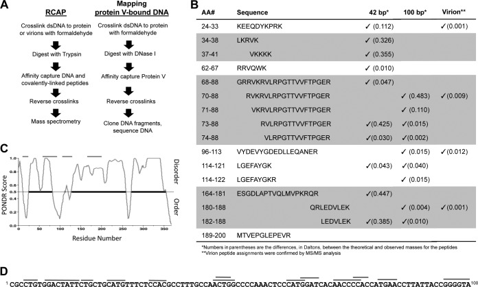
FIG 4.
Mapping the DNA binding sites on protein V. (A) Scheme used for the reversible cross-linking affinity purification protocol (see Materials and Methods for details) and for the identification of DNA sequences that contact protein V. (B) Summary of the results from MALDI-TOF mass spectra of peptides that cross-linked to dsDNA of 42 bp or to the genomic DNA within the virion. Peptides that are overlapping are shown in the regions of the figure colored gray. (C) Plot of protein V intrinsic disorder prediction. The prediction was made using PONDR-FIT, version VL-XT (55). The upper gray lines show the peptides identified in the RCAP assay superimposed on the regions predicted to have secondary structure or intrinsic disorder. The heavy black lines represent portions of protein V predicted to be intrinsically disordered. (D) Mapping of the sequences in a 100-bp DNA that contacts protein V. DNA fragments that bind protein V are identified by the gray lines.

