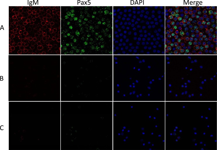
FIG 3.
Confocal micrographs of koi peripheral WBC. IgM+ cells were identified by anti-carp IgM monoclonal antibody and secondary goat anti-mouse IgG coupled with Texas Red (red). Pax5+ cells were identified by anti-Pax5 polyclonal antibody and secondary goat anti-rabbit IgG antibody coupled with Alexa Fluor 488 (green). The nucleus was identified with DAPI (blue). The Merge images shows cells visualized by all three staining methods. (A) IgM+ WBC selected by magnetic column; (B) IgM− nonselected cells that passed through magnetic column; (C) peripheral WBC before the cells were sorted on the magnetic column.

