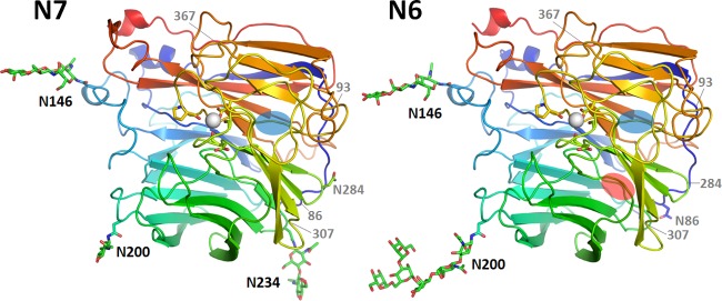
FIG 1.

Overall structures of N7 and N6. N7 (left) adopts a propeller-like structure whereby each monomer has six β-sheets (named blades 1 to 6), with blade 6 containing only three β-strands. Each N6 monomer (right) has six blades, with blades 4 and 6 containing only three β-strands. The missing β-sheets in blade 6 and blade 4 are indicated by blue ovals and a red oval, respectively. Three N-glycosylation sites, Asn146, Asn200, and Asn234, are observed in N7, and two N-glycosylation sites, Asn146 and Asn200, are observed in N6. These occupied N-glycosylation sites are indicated by black labels, the putative N-glycosylation sites with no observed glycans are labeled with an N followed by the position number, and N-glycosylation sites that are present in other influenza virus NA structures are indicated by gray labels with only the position number. There is a single calcium ion (white sphere) binding site in each N7 and N6 monomer. In both structures, the calcium ion is coordinated by Asp293, Gly297, Asp324, and Pro347.
