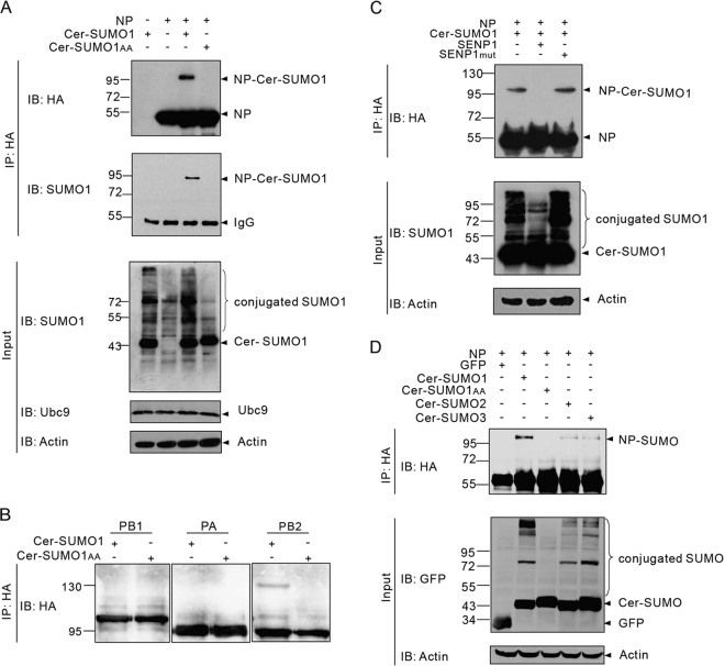
FIG 1.
Sumoylation of NP in vivo. (A) 293T cells were transfected with plasmids expressing WSN NP and Ubc9 together with plasmids expressing either Cer-SUMO1 or Cer-SUMO1AA, as indicated. The cells were lysed at 36 h posttransfection, and NP was immunoprecipitated (IP) with anti-HA–agarose. The NPs were immunoblotted (IB) with anti-HA polyclonal antibodies. Cer-SUMO1 was detected in separate blots with an anti-SUMO1 monoclonal antibody. (B) 293T cells were transfected with plasmids expressing an HA-tagged WSN polymerase protein (PB1, PA, or PB2) and Ubc9 together with plasmids expressing either Cer-SUMO1 or Cer-SUMO1AA. The cells were lysed at 36 h posttransfection, and each polymerase protein was immunoprecipitated with anti-HA–agarose and further analyzed by SDS-PAGE and immunoblotting with anti-HA polyclonal antibodies. (C) 293T cells were transfected with WSN NP and Ubc9 together with Cer-SUMO1, SENP1, or SENP1mut, as indicated. NP was immunoprecipitated with anti-HA–agarose, and the precipitated proteins were further analyzed by SDS-PAGE and immunoblotting with anti-HA polyclonal antibodies. (D) WSN NP and Ubc9 were cotransfected with GFP (as a control), Cer-SUMO1, Cer-SUMO1AA, Cer-SUMO2, and Cer-SUMO3, as indicated; NP was immunoprecipitated with anti-HA–agarose, and the precipitated proteins were further analyzed by SDS-PAGE and immunoblotting with anti-HA polyclonal antibodies. Input of the whole-cell lysates was detected by immunoblotting with anti-GFP polyclonal antibodies to show the expression of GFP, Cer-SUMO1, Cer-SUMO2, or Cer-SUMO3; with anti-Ubc9 antibody to show the expression of Ubc9; and with anti-actin antibodies to show the loading controls. The values to the left of the blots are molecular sizes in kilodaltons.

