FIG 5.
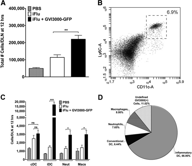
GVI3000 enhances inflammation in the DLNs of neonatal mice. Five 7-day-old BALB/c mice were immunized in both f.p. with PBS only or with iFlu (1 μg) in the presence of 105 IU of VEE replicon particles labeled with GFP (VRP-GFP) or absence of VRP-GFP. At 12 hpi, both popliteal DLNs from each neonatal mouse were harvested, combined, and manually disrupted into a single-cell suspension. (A) DLN cellularity was assessed by using the volume analyzed by an Accuri C6 flow cytometer (BD) and back-calculating the starting sample amount. (B) Representative histogram for inflammatory dendritic cell gating. (C) Using fluorescent antibody staining, immune cells were stained for DCs (CD11c+), inflammatory DCs (CD11c+ Ly6Chi), neutrophils (Ly6G+), and macrophages (CD11c− CD11b+). The total number of each immune cell present in the DLN sample is shown in the graph. Neut, neutrophils; Macs, macrophages. (D) Cells that were positive for GFP expression were then gated for the surface markers of different immune cells. In panels A and C, the data are presented as means plus SEM. Values that are significantly different by the Mann-Whitney test are indicated by bars and asterisks as follows: *, P < 0.05; **, P < 0.01; ***, P < 0.001. Values that are not significantly different (ns) are indicated.

