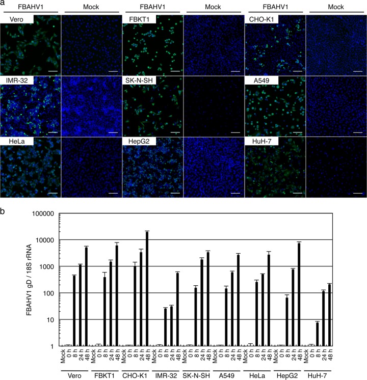
FIG 4.
FBAHV1 infection of different cell types. (a) Each cell line was infected with FBAHV1 (left) or mock infected (right) for 16 h at an MOI of 0.1. After fixation, FBAHV1 antigens (green) were visualized by staining with an anti-HSV-1 polyclonal antibody, followed by Alexa Fluor 488-conjugated anti-rabbit IgG. Cell nuclei (blue) were stained with Hoechst 33342. All cell lines tested were susceptible to FBAHV1 infection. Bars, 100 μm. (b) DNA was extracted from FBAHV1-infected cells (MOI = 0.5) at the indicated times p.i. FBAHV1 genomic DNA was quantified by real-time PCR, and the amount was normalized to that of 18S rRNA. All cell lines tested supported virus replication. Data are expressed as the mean ± standard error of the mean (SEM) from triplicate reactions.

