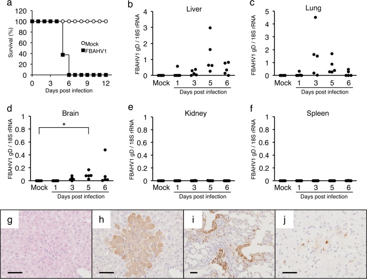
FIG 5.
Experimental infection of laboratory mice with FBAHV1. (a) BALB/c mice were either infected intranasally with 105 PFU of FBAHV1 (n = 8) or mock infected (n = 4). Survival was monitored daily for 12 days. (b to f) Tissues were harvested from five FBAHV1-infected mice at the indicated time points. The amount of the FBAHV1 genome in tissues was determined by real-time PCR and normalized to the amount of 18S rRNA. Four mock-infected mice were included in the analysis as negative controls. *, P < 0.05. (g) Hematoxylin-eosin staining of liver tissue sections from mice at 5 days postinfection with FBAHV1. (h to j) Immunohistochemical staining of liver (h), lung (i), and brain (j) tissues from mice at 5 days postinfection with FBAHV1. FBAHV1 antigens (brown) were detected by staining with an anti-HSV-1 polyclonal antibody. Bars, 50 μm.

