
FIG 4.
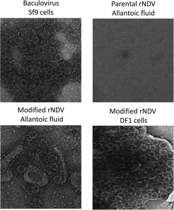
Production of VLPs by rNDV. Shown are electron microscopy images of norovirus VLPs. VLP suspensions (10 μl each) were fixed in copper grids, negatively stained with 1% ammonium molybdate, and visualized by using an electron microscope.
Official websites use .gov
A
.gov website belongs to an official
government organization in the United States.
Secure .gov websites use HTTPS
A lock (
) or https:// means you've safely
connected to the .gov website. Share sensitive
information only on official, secure websites.
Production of VLPs by rNDV. Shown are electron microscopy images of norovirus VLPs. VLP suspensions (10 μl each) were fixed in copper grids, negatively stained with 1% ammonium molybdate, and visualized by using an electron microscope.