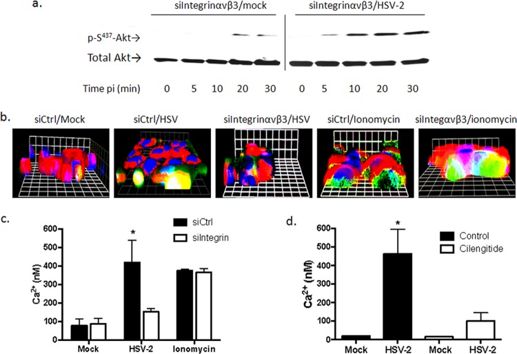
FIG 3.
Integrin αvβ3 contributes to the virus-induced cytoplasmic Ca2+ response post-Akt phosphorylation. (a) CaSki cells transfected with siIntegrinαvβ3 RNA were mock infected (serum free medium) or infected with purified HSV-2(G) (10 PFU/cell), and cell lysates were prepared for Western blot analysis at the indicated times p.i. Blots were incubated with anti pS473-Akt123 and then stripped and probed with anti-total Akt123. A representative blot from 3 independent experiments is shown. (b) CaSki cells were loaded with Calcium Green 72 h posttransfection with the indicated siRNA and synchronously mock infected or infected with purified HSV-2(G) (5 PFU/cell). Live images were acquired 3 min after a temperature shift to 37°C. Cellular membranes were stained with CellTrace (red), nuclei were stained with Hoechst (blue), and Ca2+ is green. Representative XYZ images from 3 independent experiments are shown. Bars = 9.2 μm. (c) Transfected CaSki cells were loaded with fura-2, infected with purified HSV-2(G) (2 PFU/cell), or mock infected. To assess whether siRNA transfections impacted the intracellular Ca2+ stores, uninfected cells were treated with 1 μM ionomycin. The mean intracellular Ca2+ concentration (nM) over 1 h was calculated from 4 wells, each containing 5 × 10⁴ cells; the asterisk indicates a significant increase in Ca2+ concentration relative to the mock-infected control (P < 0.05). (d) Parallel studies were conducted with cells treated with calcium-free buffer (control) or buffer supplemented with cilengitide (100 μM), and the Ca2+ response over 1 h p.i. was compared to that of mock-infected cells.

