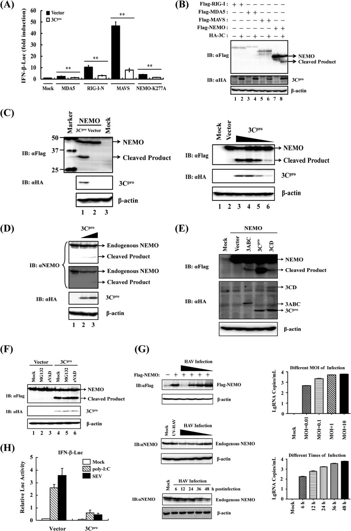
FIG 2.
HAV 3Cpro disrupts RLR signaling by cleaving NEMO. (A) HEK293T cells cultured in 24-well plates were cotransfected with IFN-β-Luc, pRL-TK plasmid, and plasmid encoding 3Cpro (0.3 μg), together with the MDA5, RIG-I-N, MAVS, or NEMO-K277A expression vector (0.7 μg). Luciferase assays were performed 36 h after transfection. **, P < 0.01 (considered highly significant). (B) HEK293T cells cultured in 60-mm-diameter dishes were transfected with Flag-tagged RIG-I, MDA5, MAVS, or NEMO expression plasmid (4 μg) along with empty vector or plasmid encoding hemagglutinin (HA)-tagged 3Cpro (0.5 μg). Cell lysates were prepared 30 h posttransfection and analyzed by Western blotting (immunoblotting [IB]). (C) (Left) HEK293T cells cultured in 60-mm-diameter dishes were transfected with Flag-tagged NEMO expression plasmid (2 μg) along with empty vector or plasmid encoding HA-tagged 3Cpro (1 μg). Cell lysates were prepared 30 h posttransfection and analyzed by Western blotting. The lane with protein markers (Bio-Rad, catalog no. 161-0376) includes 50-, 37-, and 25-kDa molecular mass bands. (Right) HEK293T cells cultured in 60-mm dishes were transfected with Flag-tagged NEMO expression plasmid (2 μg), along with increasing quantities (0, 0.125, 0.25, 0.5, or 1 μg) of plasmid encoding HA-tagged 3Cpro. Cell lysates were prepared 30 h posttransfection and analyzed by Western blotting. (D) HEK293T cells cultured in 60-mm dishes were transfected with increasing quantities (0, 2, or 4 μg) of plasmid encoding HA-tagged 3Cpro. Cells were lysed 30 h posttransfection and analyzed by Western blotting. (Two different exposures of the blot are shown.) (E) HEK293T cells cultured in 60-mm dishes were transfected with Flag-tagged wild-type NEMO as indicated (2 μg), along with HA-3Cpro or 3Cpro-containing precursors (1 μg). Cell lysates were prepared 30 h posttransfection and analyzed by Western blotting. (F) HEK293T cells cultured in 60-mm dishes were cotransfected with Flag-tagged NEMO expression plasmid (2 μg) and plasmid encoding 3Cpro or empty vector (1 μg). Twenty-four hours after transfection, MG132 or zVAD-FMK was added to a final concentration of 20 μM. Cell lysates were prepared 8 h after treatment and analyzed by Western blotting. (G) (Top) HEK293T cells cultured in 60-mm dishes were transfected with Flag-tagged NEMO (Flag-NEMO) and infected with different doses (multiplicities of infection [MOI] of 0.01, 0.1, 1, and 10) of HAV (L-A-1 attenuated vaccine strain) 6 h posttransfection. The cells were lysed 36 h postinfection and analyzed by Western blotting. (Middle) HEK293T cells were infected with different doses of HAV or, as a control, UV light-inactivated HAV (UV-HAV). The cells were lysed 36 h postinfection and analyzed by Western blotting. (Bottom) HEK293T cells were infected with HAV (MOI of 1), lysed at different times of HAV postinfection, and analyzed by Western blotting. Expression of Flag-NEMO conjugated protein and endogenous NEMO protein was verified by mouse anti-Flag antibody (Macgene, China) or rabbit anti-NEMO (Abclonal, China), respectively. Mouse anti-HA (Abclonal, China) was used to confirm the expression of 3Cpro, and mouse anti-β-actin antibody (Beyotime, China) was used to detect β-actin, which serves as a protein loading control. (H) Activation of the IFN-β promoter by extracellular poly(I·C) (20 μg/ml) or SEV (100 hemagglutinating units [HAU]/ml) in HEK293-TLR3 cells transiently expressing 3Cpro or a control vector.

