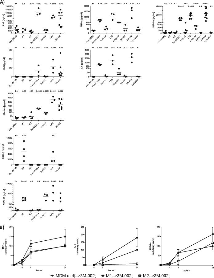
FIG 4.
The most prominent cytokine secretion in response to TLR triggering was in M1-polarized macrophages, followed by MDMs. (A) MDMs were polarized or exposed to Pam2CSK4, poly(I·C), LPS, flagellin, 3M-001, 3M-002, and CpG2006 for 24 h, and supernatants were analyzed for cytokines and chemokines (n ≥ 3). (B) M1- and M2-polarized macrophages were challenged with 3M-002 (TLR8 agonist) for 24 h. Subsequently, supernatants were collected to quantify the amounts of TNF-α, IL-6, and macrophage inflammatory protein 1α (MIP-1α) released (n = 3). MDM (ctrl) → 3M-002, untreated macrophages (control) treated with 3M-002; M1 → 3M-002, M1-polarized macrophages treated with 3M-002; M2 → 3M-002, M2-polarized macrophages treated with 3M-002. The agonists are described in the legend to Fig. 1. The results for MDMs (control [Ctrl]) and either polarized macrophages or macrophages treated with the distinct TLR agonists were compared by the unpaired t test with a one-tailed P value.

