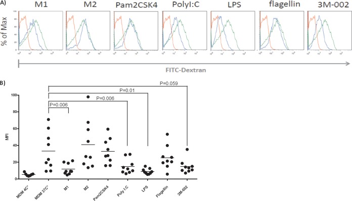
FIG 7.
M1 polarization and triggering of TLR3, -4, and -8 result in substantially less endocytic activity than that from MDMs. For assessing endocytic activity, polarized macrophages or TLR-primed MDMs were incubated with 0.1 mg/ml FITC-dextran of 70 kDa for 1 h and harvested to quantify macrophages that had taken up FITC-dextran by flow cytometry. (A) Mean fluorescence intensity of one representative example. Red lines, macrophages exposed to FITC dextran at 4°C; green lines, MDMs; blue lines, MDMs treated with the various TLRs. (B) Compilation of the results of all the experiments performed. Data from assays with FITC-dextran at either 10 or 150 kDa were similar. We used the paired t test to calculate the statistics. The agonists are described in the legend to Fig. 1. MFI, mean fluorescence intensity.

