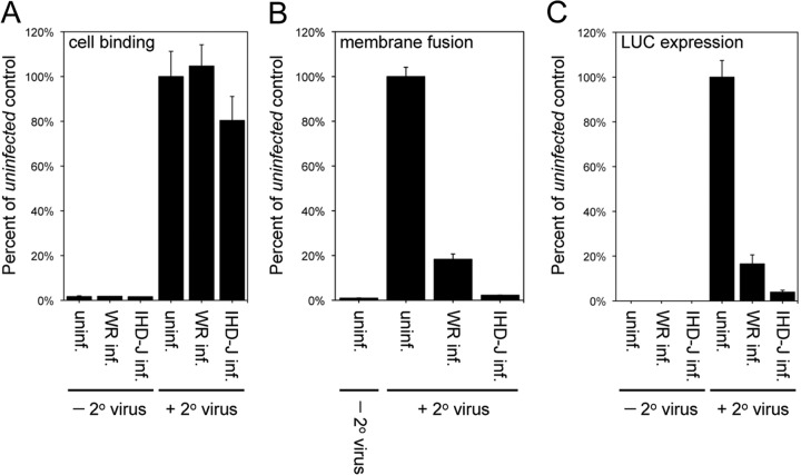
FIG 3.
SIE occurs at the virus-cell membrane fusion step. (A) Cell binding step. Cells were left uninfected or infected with 10 PFU/cell of VACV WR or IHD-J for 120 min and incubated with 5 PFU/cell of YFP-tagged secondary VACV WR at 4°C for 60 min. Cells were analyzed by flow cytometry to determine the number and mean fluorescence of YFP+ cells. Data are presented as the percentage of YFP-tagged virus bound to primary virus-infected cells compared to uninfected control cells. (B) Hemifusion step. Cells were uninfected or infected with 10 PFU/cell of VACV WR or IHD-J for 120 min. The cells were superinfected with 3 PFU/cell of DiD-loaded secondary VACV WR at 37°C for 90 min and analyzed by flow cytometry to determine the number of DiD+ cells and the DiD mean fluorescence intensity. The latter values were normalized to the values obtained for the uninfected control cells infected with DiD-loaded virus. (C) LUC expression. Cells were uninfected or infected with 10 PFU/cell of VACV WR or IHD-J for 120 min and superinfected with 3 PFU/cell of WRvFire for 150 min. Cells then were lysed and LUC activity quantified; the data were normalized to values obtained for the uninfected control cells infected with WRvFire.

