
FIG 5.
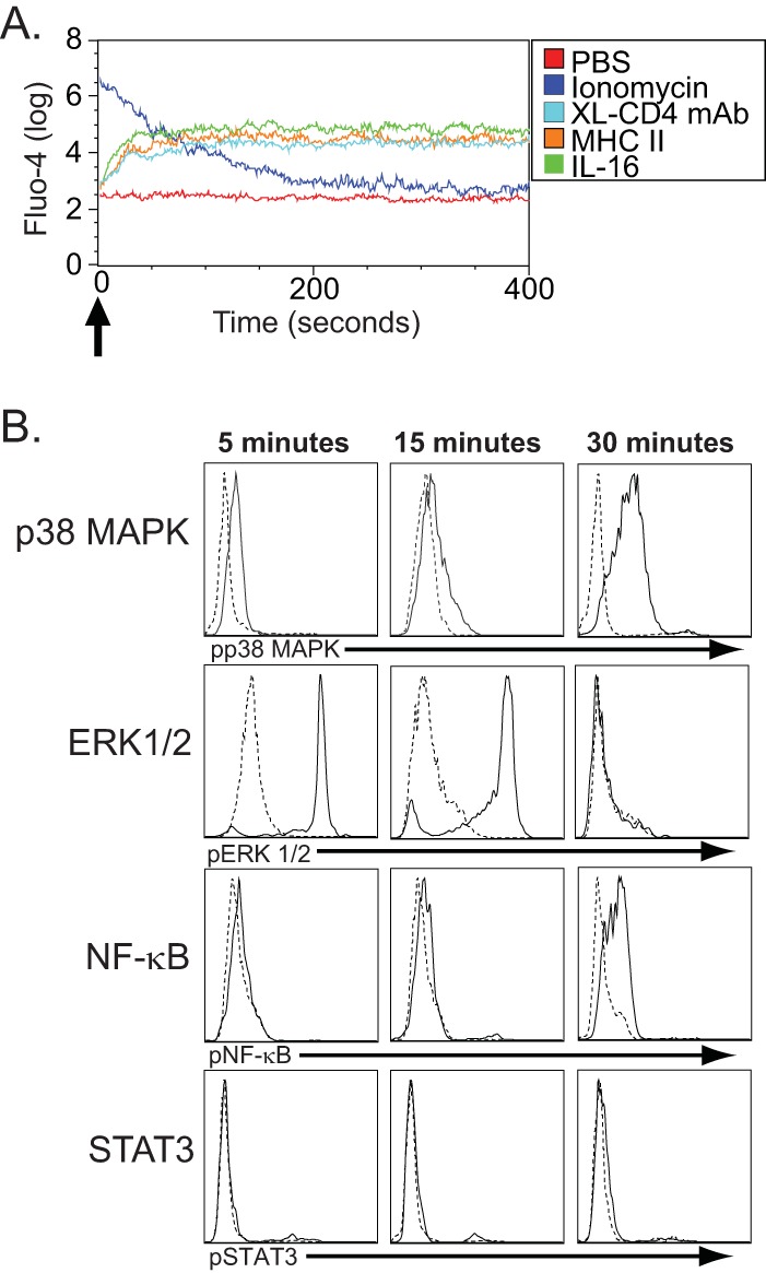
CD4-mediated signal transduction in negatively selected fresh human monocytes. (A) Ca2+ flux following CD4 ligation on human monocytes. Freshly isolated human monocytes were labeled with Fluo-4 and then treated (indicated by the arrow) with either PBS alone (negative control), ionomycin (positive control), cross-linked CD4-specific monoclonal antibodies (XL-CD4 MAb), sMHC-II, or IL-16. They were immediately measured for Ca2+ flux by flow cytometry and analyzed for fluorescence of Fluo-4 (y axis) for the indicated time (x axis) following stimulation. (B) Phosphorylated p38 MAP kinase (pp38MAPK), ERK1/2 (pERK1/2), NF-κB (pNF-κB), and STAT3 (pSTAT3) were assessed in cells treated with medium only (dashed lines) and cells treated with sMHC-II (solid lines) 5, 15, and 30 min following stimulation. The histogram depicts the relative fluorescence intensities, as determined by flow cytometry, for cells staining for pp38MAPK, pERK1/2, pNF-κB, and pSTAT3 and is representative of 3 separate experiments.