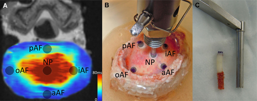
Figure 2.
IVD test sites. (A) Site locations on a transverse slice of a quantitative T2* map. The average T2* relaxation time was measured at each site. (B) Same test locations on a specimen undergoing stress relaxation tests using hybrid confined/in situ indentation methodology to compute the residual stress and excised strain of the tissue. (C) A 3 mm punch was used to remove a plug of tissue at the same locations where indentation tests were performed and where MRI relaxation times were recorded. The endplate and inferior trabecular bone were removed prior to biochemical quantification. In a randomized fashion, the superior or inferior section was used to quantify either s-GAG and water content or hydroxyproline content.

