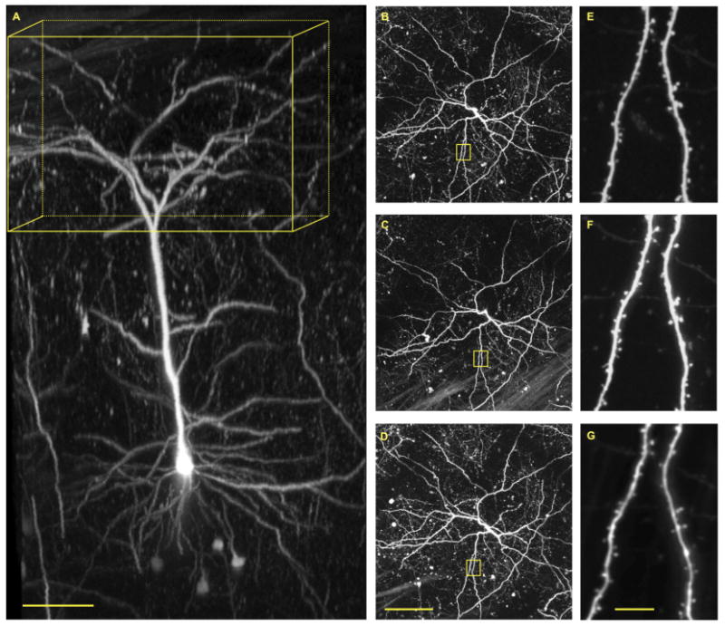
Fig. 8.

Repetitive imaging through a cranial window of M-line male mouse. (A) Three-dimensional reconstruction of a single layer 5 pyramidal neuron in the sommatosensory cortex. Yellow box has dimensions of 400 x 400 x 200 microns (x/y/z). (B) Maximum intensity projections of the boxed volume in A. (C) Maximum intensity projection of the boxed volume in A taken 7 days after the first image. (D) Maximum intensity projection of the boxed volume in A taken 21 days after the first image. (E-G) Magnified images of dendritic tree in yellow squares in B-D. Cellular labeling in M-line male mice with eGFP is so sparse (see Figure 3) and intense that it allows facile two-photon fluorescence imaging of single neurons with great clarity. Note, basal dendrites are visible at a depth of 500-600 microns below the pia. Repetitive images of the entire neuron revealed the overall structure of the dendritic tree is very stable (B-D). High-resolution imaging revealed most spine heads are also stable over a 21-day period (E-G). The window was implanted when the mouse was 121 days old, and imaging commenced at 62 days later. Scale bars: 100 microns (A,D) 100 microns; G 10 microns.
