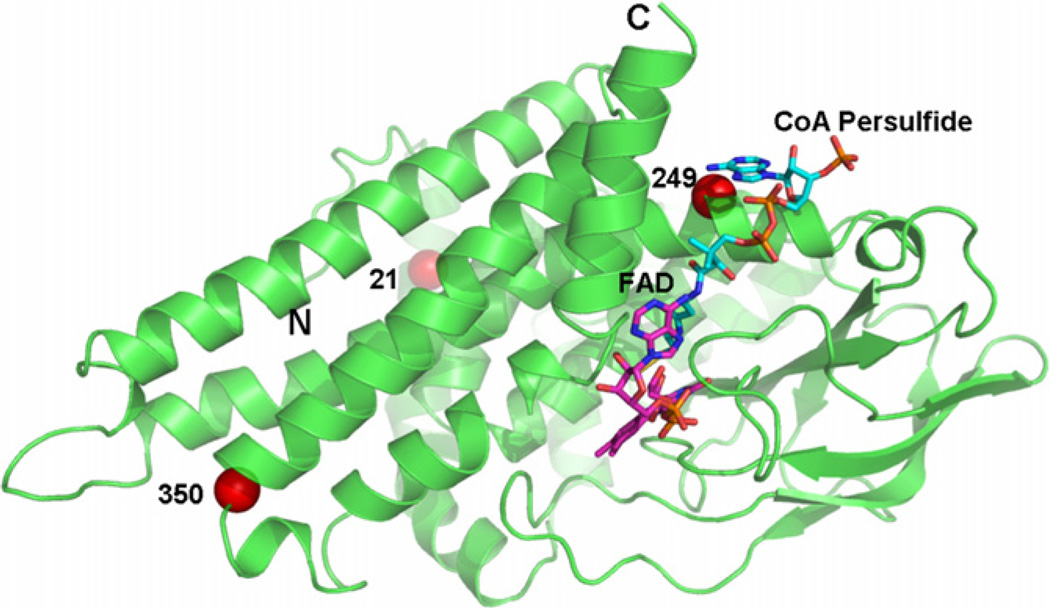
Fig. 3.
Ribbon diagram of a monomer of human IVD with bound CoA persulfide depicting the locations of the amino acid replacements identified in Korean IVA patients. Substituted amino acid residues are denoted with red-colored balls. (For interpretation of the references to colour in this figure legend, the reader is referred to the web version of this article.)

