
Figure 2. Use of automated bright-field imaging (ImageXpress) for in vivo screening.
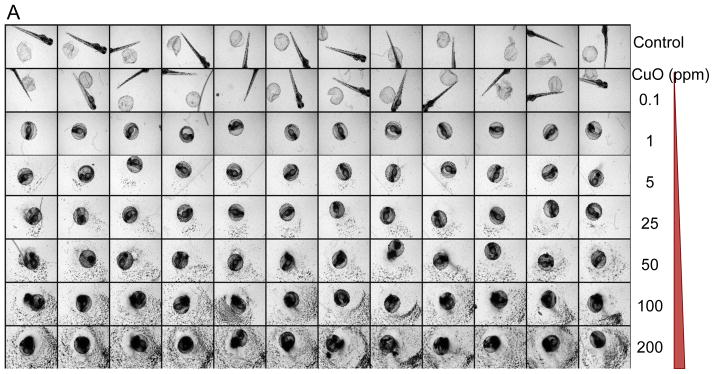

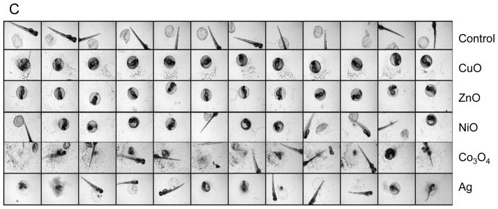

(A) A representative image of a 96-well plate containing embryos viewed at 72 hpf in the presence of CuO nanoparticles. Each embryo was hand picked and placed in the well of a multiwell plate, which subsequently received 0.1–200 ppm CuO nanoparticles, starting at 4 hpf. Bright-field phase contrast images of each well were obtained through a 2x dry objective that auto-focuses on each plate at 24 hr intervals for 5 consecutive days. Each treatment group, including non-exposed controls, was assessed using 12 replicates. (B) The survival and hatching rates of embryos at 72 hpf clearly demonstrate hatching interference by CuO nanoparticles. Although the hatching interference was profound at doses ≥ 0.5 ppm, there was no apparent adverse effect on embryo survival, even at the highest CuO nanoparticle concentration (200 ppm). The statistical calculations are based on cumulative screening of 2000 embryos. Our high content capacity now enables us to screen 400 embryos every 72 hours, amounting to 800 embryos per week. (C) A representative image of a 96-well plate comparing the screening of CuO, ZnO, NiO, Co3O4, and Ag nanoparticles (positive control) at 50 ppm. Parallel comparison across different nanoparticle types was accomplished using the same high content imaging technique. (D) Statistical analysis of embryos survival and hatching rates. Statistical significance was determined as p<0.05 according to the student t-test.