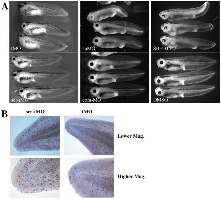
Figure 5. Effect of xGDF11 knockdown on embryonic phenotype.
(A) Phenotype of stage 41 embryos injected with 60 ng tMO versus scr-tMO (left), and of stage 40 embryos injected with 20 ng spMO or cont-MO (center), and stage 40 embryos treated with 100 mM SB-431542 or 0.1% DMSO at stage 13. B) Cell proliferation was measured by phospho-histone H3 staining of tails from at Stage 40 embryos treated with 60 ng xGDF11 tMO (right) or 60 ng xGDF11 scr-tMO left. High magnification is of each tail tip is shown at bottom. Data shown are typical of three separate experiments, a similar reduction in proliferation with xGDF11 knockdown was seen when measured using BrdU incorporation (not shown).

