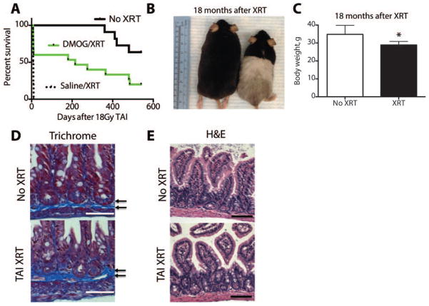
Figure 7. Long-term survivors of lethal TAI exhibit lower body weights and microscopic intestinal changes.
(A) Kaplan Meier analysis of mice treated with saline, DMOG or no XRT 18 months after 18Gy TAI. Log rank test showed p=0.01 XRT vs No XRT. (B) Surviving mice from the DMOG cohort treated with TAI (right) and an age matched control (left). Note the change in fur color in the irradiated lower body. (C) Body weight of mice at approximately 20 months of age. (*p=0.02, n=8/group). (D) Trichrome and (E) H&E stains from intestines from mice 18 months after receiving TAI or no radiation. Arrows indicate microscopic fibrotic bands. Scale bars=50μm.

