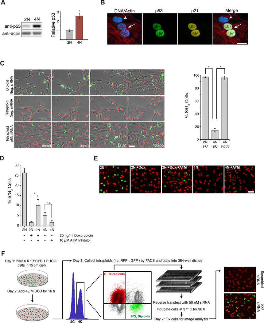
Figure 1. Genes that mediate cell cycle arrest of tetraploid cells.
(A) Western blot of p53 levels in diploid (2N) and tetraploid (4N) cells 48 h after sorting (n=6; *p < 0.001, unpaired t-test). (B) Diploid (arrowhead) and binucleated tetraploid (arrow) cells stained for p53 (green), p21 (yellow), actin (red), and DNA (blue). Scale bar, 25 µm. (C) Still images of sorted diploid and tetraploid RPE-FUCCI cells that were transfected with the indicated siRNAs (from a live-cell imaging experiment). The percentage of cells that progress into S-phase from 5 independent imaging experiments is shown on right (2N, n=119; 4N, n=326; 4N p53 siRNA, n=211; *p < 0.00001, unpaired t-test). Time, hrs:mins. Scale bar, 100 pm. (D) The fraction of S/G2 cells from diploid (2N) and tetraploid (4N) cells following treatment with ± 25 ng/ml doxorubicin and ± 10 µM ATM inhibitor for 24 h (n=3; *p < 0.05, unpaired t-test, n.s, not significant). (E) Representative images of FUCCI cells from the experiment in (D). Scale bar, 100 µm. (F) Protocol for genome-wide RNAi screen to identify genes necessary to activate or maintain G1 cell cycle arrest in tetraploid cells after cytokinesis failure. All error bars represent mean ± SEM.

