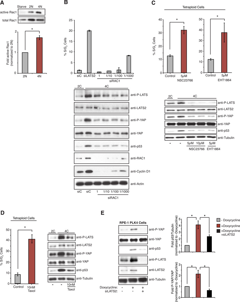
Figure 5. Increased Rac activity triggered by extra centrosomes activates the Hippo pathway.
(A) Western blot analysis and quantitation of pull-down assays to measure active Rac1 relative to total Rac1 in serum-starved, 2N, and 4N RPE-1 cells (n=5; *p < 0.003, one sample t-test). (B, C, D) The percentage of S/G2 tetraploid RPE-FUCCI cells following siRNA-mediated depletion of Rac1 (B), ± 5 µM treatment with the Rac inhibitors NSC2376 and EHT1864 (C), or 10 nM treatment with Taxol (D), along with corresponding western blot analysis of Hippo pathway activation (n ≥ 3; *p < 0.01, unpaired t-test). (E) Western blot analysis (left) and quantitation (right) of Hippo activity and p53 levels in RPE-1 cells with or without transient doxycycline-induced PLK4 overexpression and extra centrosomes. Cells were treated with control or LATS2 siRNAs prior to doxycycline treatment (n=10; *p < 0.01, unpaired t-test). Error bars represent the mean ± SEM.

