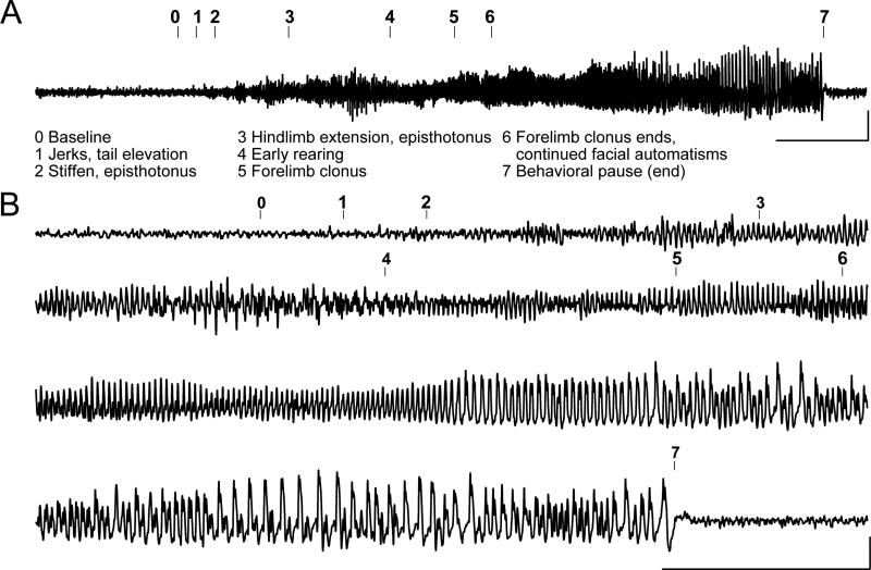
Figure 3. R6/2 mice experience spontaneous electrographic seizures with corresponding seizure behaviors.
(A) Representative compressed electroencephalogram from cortical lead depicting a spontaneous electrographic seizure in an R6/2 mouse, with seizure behaviors identified at the time of occurrence (numbers). Background EEG is shown before seizure onset, evolves into rhythmic, sharply contoured spikes, and resolves with clear postictal suppression accompanied by a behavioral pause; calibrator depicts 0.0005V, 20s. (B) Time expanded EEG recording calibrator depicts 5s, 0.0005V.

