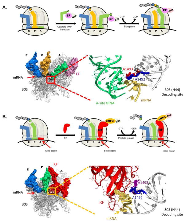
Figure 8.
Schematic view of translation elongation and translation termination with magnified elements from ribosome crystal structures. (A) Molecular glance at the bacterial decoding site upon cognate tRNA binding – 'ON' state. The 30S ribosomal subunit is showed at the left side with three tRNA molecules bound (A-site – green; P-site – Yellow; E-site – Blue) and mRNA highlighted in black. The A-site tRNA is bound to an EF (elongation factor – light pink) which is an intrinsic protein participating in the translation elongation process. The actual decoding site is highlighted in red, and is enlarged in the right side of the figure. The mRNA in the enlarged view is highlighted in yellow; A-site bound tRNA in green; and the two conserved adenine residues, A1492 and A1493, flipping out from the helical core, are highlighted in blue and red, respectively. The PDB entry for the presented structure is 2WRQ. (B) Structural view of the bacterial decoding site during termination, with mRNA highlighted in black; P-site and E-site tRNA molecules highlighted in green and blue, respectively; and RF highlighted in red. The decoding site is highlighted in yellow and an enlarged representation is presented in the right side of the figure. In the enlarged object RF is highlighted in red; mRNA in gold; and the conserved adenine residues A1492 and A1493 in blue and purple, respectively. The PDB entry for the presented structure is 2X9R.

