Figure 1.

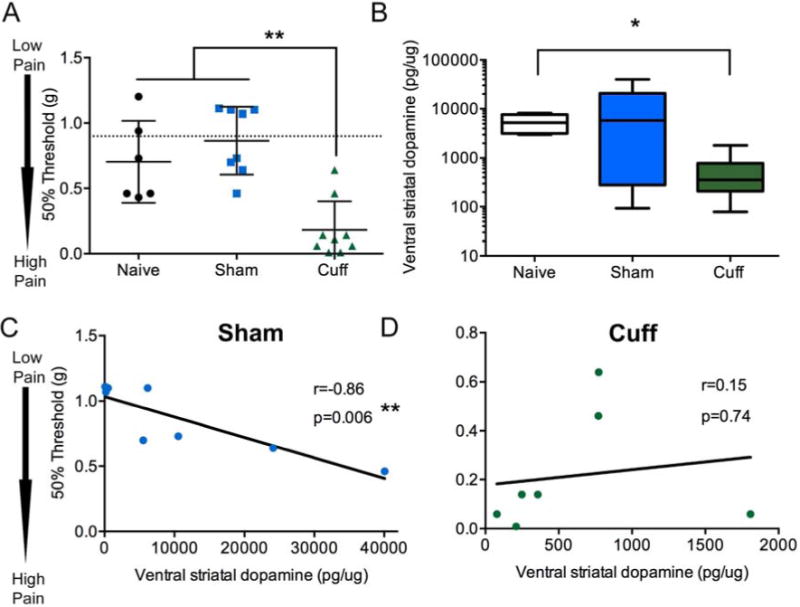
Total dopamine (DA) content in the ventral striatum increase in the cuff group, but lose correlation with mechanical thresholds. A) Two weeks following nerve injury, mechanical thresholds of the hindpaw, as measured with von Frey filaments, were significantly lowered in the cuff group. Naïve and sham groups did not have significantly different thresholds from baseline (pre-surgery, indicated by dotted line). Data represented as scatter plot. Horizontal bar represents mean and error bars represent standard error (S.E.M). Groups were compared using a one-way ANOVA followed by Tukey’s post hoc analysis. **=p<0.01, n=6–8. B) Dopamine content normalized to total protein in ventral striatum was measured by HPLC. Two weeks after nerve injury, cuff animals had significantly lowered dopamine content compared to naïve group. Sham animals were not significantly different, although had a larger variability. Data represented as box plot with horizontal line representing the mean, the outer box limits representing the interquartile span, and the whiskers the furthest data point. Groups were compared with a Kruskal-Wallis ANOVA followed by Dunn’s multiple comparison post hoc analysis. *=p<0.05, n=6–8. C) Dopamine content normalized to total protein in the ventral striatum were correlated with mechanical thresholds 2 weeks after nerve injury. The sham group showed significant negative correlation between dopamine and mechanical thresholds, while the (D) cuff group failed to show this correlation. Correlation was performed with Pearson’s correlation. n=7–8.
