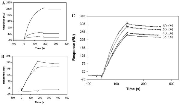
Fig. 1.
(A and B) SPR sensorgrams of nonspecific binding of factor P in PBS (A) and HBS-EP (B) with (1) dextran (CM5 chip), (2) flat gold surface (Au sensor chip), (3) mPEG (mPEG sensor chip). (C) SPR sensorgrams (black lines) of the interaction of factor P with heparin. Gray lines represent the theoretical curves obtained from a global fitting of the sensorgrams using a Langmuir 1:1 binding model with mass transport limitation. The low density of bound heparin limits the sensitivity of SPR to 35 nM of factor P. After 1200 s of dissociation, 15% of the factor P–heparin complex is dissociated.

