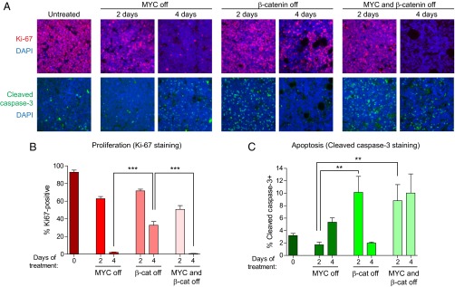
Fig. 6.

Combined inactivation of MYC and β-catenin synergizes to inhibit proliferation and promote apoptosis in vivo. (A) MYC lymphoma cells expressing DD-ICAT were transplanted s.c. into recipient SCID mice. Mice were either left untreated or treated with DOX (MYC off), TMP (β-catenin off), or both DOX and TMP (MYC and β-catenin off) for 2–4 d. Tumors were harvested and stained for the proliferation marker Ki-67 and the apoptotic marker cleaved caspase-3. Nuclei were stained using DAPI. Representative images are shown. (B and C) Quantification of Ki-67 and cleaved caspase-3 staining, in two to three independent tumors from each time point and five fields per tumor. Values represent means ± SEM. **P < 0.01, ***P < 0.001.
