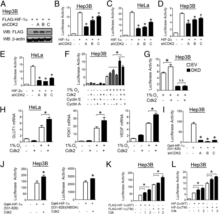
Fig. 6.
Cdk2 enhances HIF-1α transactivation function in cancer cells. (A) Hep3B cells were cotransfected with FLAG–HIF-1α vector and empty vector or vector encoding one of three shRNAs targeting Cdk2. At 48 h posttransfection, cell lysates were analyzed by WB. (B–E) Hep3B (B and D) and HeLa (C and E) cells were cotransfected with p2.1, pSV-RL, FLAG–HIF-1α vector, and the indicated shRNA vector. At 48 h posttransfection, luciferase activities were determined. (F) Hep3B cells were cotransfected with p2.1, pSV-RL, and the indicated expression vector. At 24 h posttransfection, cells were exposed to 20% or 1% O2 for an additional 24 h, and luciferase activities were determined. (G) Hep3B cells stably transfected with either shEV (white) or shRNAs against both HIF-1α and HIF-2α [double knockdown (DKD); black] were cotransfected with p2.1, pSV-RL, and Cdk2 vector as indicated. At 24 h posttransfection, cells were exposed to 20% or 1% O2 for an additional 24 h and luciferase activities were determined. (H) HeLa cells were transfected with empty vector or Cdk2 vector followed by exposure to 20% or 1% O2 for an additional 24 h, and qRT-PCR was performed. (I) Hep3B cells were cotransfected with vector encoding Gal4–HIF-1α(531–826), firefly luciferase reporter pG5E1bLuc, pSV-RL, and the indicated shRNA vector. At 48 h posttransfection, luciferase activities were determined. (J) Hep3B cells were cotransfected with vector encoding Gal4–HIF-1α(531–826) or Gal4–HIF-1α(531–826)(N803A), pG5E1bLuc, pSV-RL, and the indicated expression vector. At 48 h posttransfection, luciferase activities were determined. (K and L) Hep3B cells were cotransfected with p2.1, pSV-RL, vector encoding wild-type (WT) or triple mutant (TM) HIF-1α (K), or HIF-2α (L), and the indicated expression vector. At 24 h posttransfection, luciferase activities were determined. All results in bar graphs are presented as mean ± SEM (n = 4). *P < 0.01; #P < 0.05; n.s., not significant.

