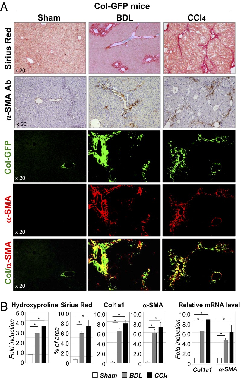
Fig. 1.
Development of liver fibrosis in Col-GFP mice in response to BDL and CCl4. (A) CCl4-treated and BDL-operated mice (but not sham mice, 8-wk-old, n = 10 per group) developed liver fibrosis, as shown by Sirius Red staining, fluorescent microscopy for collagen-GFP, and staining for α-SMA (20× objective). (B) Fibrosis was assessed by hydroxyproline and Sirius Red (positive area) content and by mRNA levels of fibrogenic genes (Col and α-SMA) in all groups of mice is shown, *P < 0.003; **P < 0.001.

