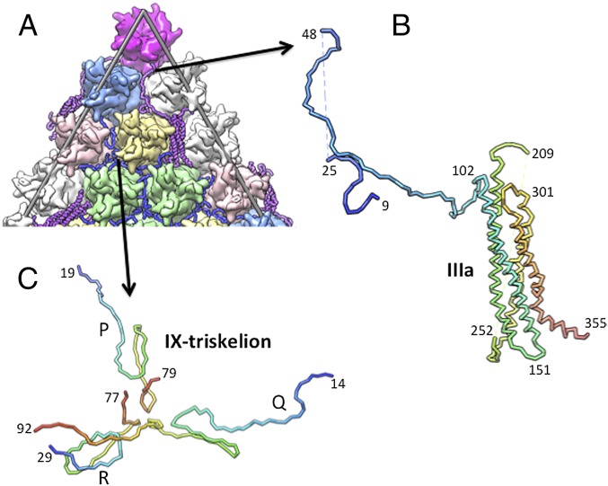
Fig. 2.
Structure and location of outer cement proteins. (A) A zoomed-in view of the (partial) icosahedral facet. (B) A tube representation showing the fold of protein IIIa. Rainbow coloring highlights the flow of the polypeptide chain from the N terminus (blue) to the C terminus (red). Dashed lines represent disordered residues. Selected residues are labeled. (C) Structure of one of the protein IX triskelions. Rainbow coloring blue to red shows the trail of the polypeptide chain from the N terminus to the C terminus for each of the three individual IX molecules P, Q, and R.

