Fig. 5.
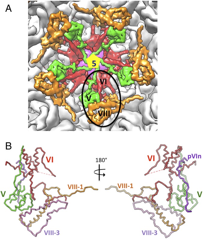
Organization of cement proteins underneath the vertex region. (A) Five copies of the ternary complex formed by proteins V (green), VI (red), and VIII (orange) glue the peripentonal hexons (gray) to each other and connect them to the adjacent GONs. One of the copies is highlighted by a black oval. (B) A stick diagram showing the structure of the ternary complex. Color assignments of the proteins are as in A, with the exception that the propeptide of VI (pVIn) is shown in purple and the C-terminal fragment of VIII (VIII-3) is shown in orchid, whereas the N-terminal fragment of VIII (VIII-1) is shown in orange.

