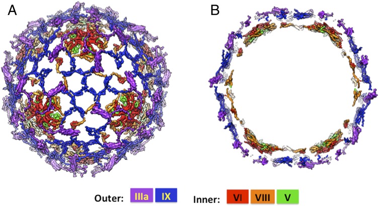
Fig. 6.
Organization of cement proteins in human adenovirus. (A) Organization of the five different minor/cement proteins IIIa (purple), IX (blue), V (green), VI (red), and VIII (orange) that are present in multiple copies. Color assignments of inner and outer cement proteins are indicated (Lower). (B) Cross-section showing the double-layered organization of the cement proteins. The outer layer is composed of proteins IIIa and IX and the inner layer is formed by proteins V, VI, and VIII. The encapsidated DNA is not ordered in the averaged electron density maps, computed from diffraction data in the resolution range 20–3.8 Å.

