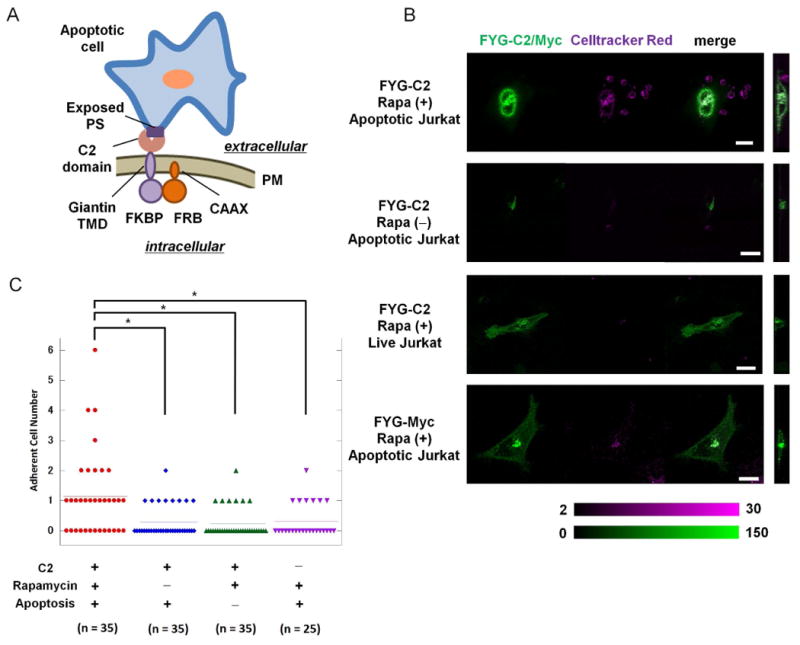
Fig. 2. Rapidly induced display of the C2 domain of MFG-E8 at the cell-surface stimulates binding to apoptotic cells.

(A) Schematic presentation of an engineered HeLa cell bound to an apoptotic Jurkat cell through the interaction between the C2 domain of MFG-E8 displayed on the surface of the HeLa cell and phosphatidylserine (PS) on the surface of the Jurkat cell. (B) HeLa cells transfected with plasmid encoding RCh-CAAX and with plasmids encoding either FYG-C2 or FYG-Myc (green) were left untreated or were treated with 200 nM rapamycin for 60 min before being incubated for 3 hours with either apoptotic or live Jurkat cells labeled with CellTracker Red (magenta). Cells were then analyzed by fluorescence microscopy to examine cell-cell adherence. Color bars indicate arbitrary fluorescence intensities for YFP (green) and CellTracker Red (magenta). Scale bar: 20 μm. Data are representative of the analysis of 25 to 35 cells from two independent experiments. (C) Scatter plots from the adhesion assays described in (B). *P < 0.05 by Mann-Whitney U-test. Each of the symbols used corresponds to the indicated experimental condition. Horizontal lines indicate the average score.
