
Figure 5.
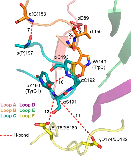
Interactions shaping the nAChR binding site, as seen in AChBP. AChBP structure and view of the binding site are identical to those in Figure 4 (PDB code 1UW6). Residue numbering is for the aligning residues of the muscle-type nAChR. Muscle-type nAChR side chains labeled are identical in AChBP, with the exceptions of G153 and P197, which are Ser and Ala, respectively, in AChBP.