
Figure 4.
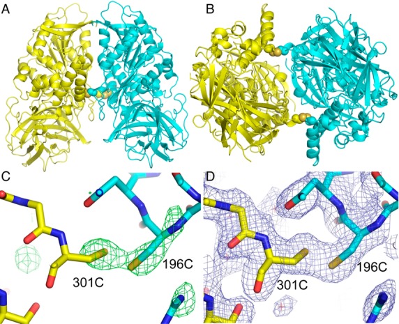
Crystal structure of the CocE mutant dimer. (A) Ribbons representation of the homodimeric molecule generated by applying 2-fold crystallographic symmetry. The side chains of the cysteine residues forming intersubunit disulfide bonds are shown in a space filling representation. (B) View of the dimer rotated 90° about a horizontal axis. (C) Fo-Fc electron density (green, 3.5 sigma contour) calculated with the rigidly placed (before refinement) molecular replacement model having residues 196 and 301 altered to glycines. The final refined model in a stick representation is superimposed on the map. (D) Final SIGMAA-weighted 2Fo-Fc electron density map (blue, 1.0 sigma cutoff) in the region of the disulfide bond with the final model shown in a stick representation.