
Figure 1.
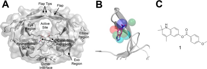
(A) Cartoon representation of HIV-1p in the semiopen conformation (PDB: 1HHP). (B) Pharmacophore model of the HIV-1p allosteric site, the Eye site, constructed by Damm et al.1 When the 5NI–protease crystal structure is superimposed on the pharmacophore model, the agreement is obvious. The pharmacophores are color-coded according to chemical property: hydrophobic (cyan), aromatic (green), hydrogen-bond donor (red), and hydrogen-bond acceptor (blue). (C) Structure of compound 1 with inhibitory activity against HIV-1p.