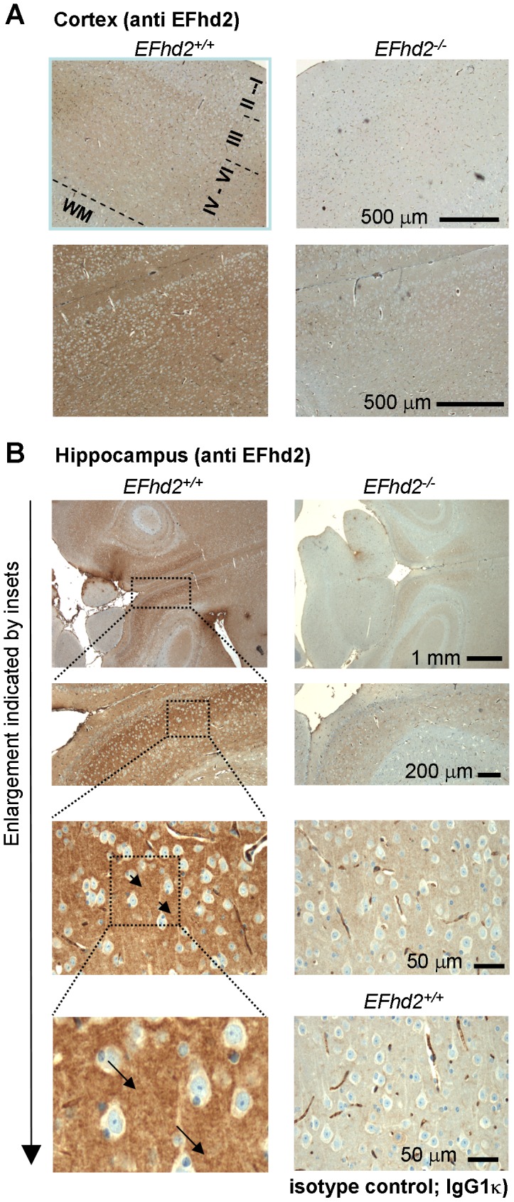
Figure 3. Specific detection of EFhd2 protein in murine brain sections.

Brains of EFhd2+/+ and EFhd2−/− mice were fixed, sectioned (horizontal) and stained with anti EFhd2 monoclonal antibody A4.18.18 in the cortex (A) or hippocampus (B), or in the hippocampus with an isotype matched control antibody (IgG1k, C). Antibodies were detected by secondary anti mouse antibody coupled to horseradish peroxidase and diaminobenzidine staining. Nuclei were counterstained with Alcian Blue. The pyramidal layers (layers III–V) and the multiform layer (layer VI) of the cortex, the molecular layer (I), the external granular layer (II) and the white matter are indicated. Representative of at least 10 mice of each genotype. Similar results were obtained with 2 additional different anti EFhd2 mAb (E7.20.23; A4.15.48) [16].
