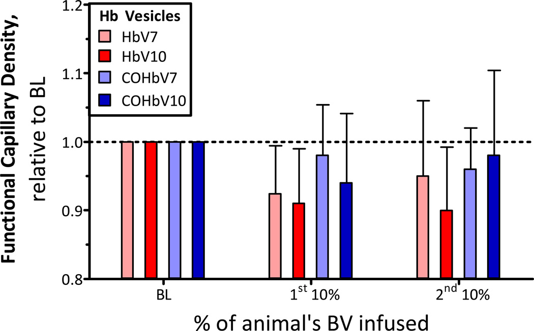
Figure 5. Relative changes from baseline in functional capillary density (FCD) after infusion of HbV or COHbV solutions.
The broken line represents the baseline level. The baseline FCD (cm−1, mean ± SD) for each group was as follows: HbV7: 110 ± 14; HbV10: 104 ± 11; COHbV7: 107 ± 13; COHbV10: 110 ± 8.

