Figure 3.
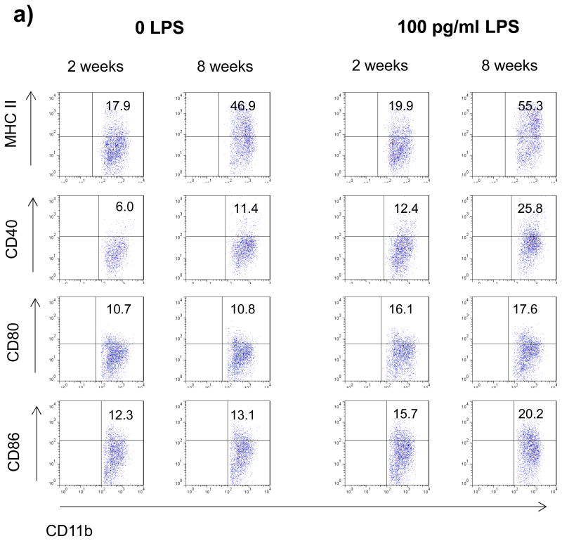
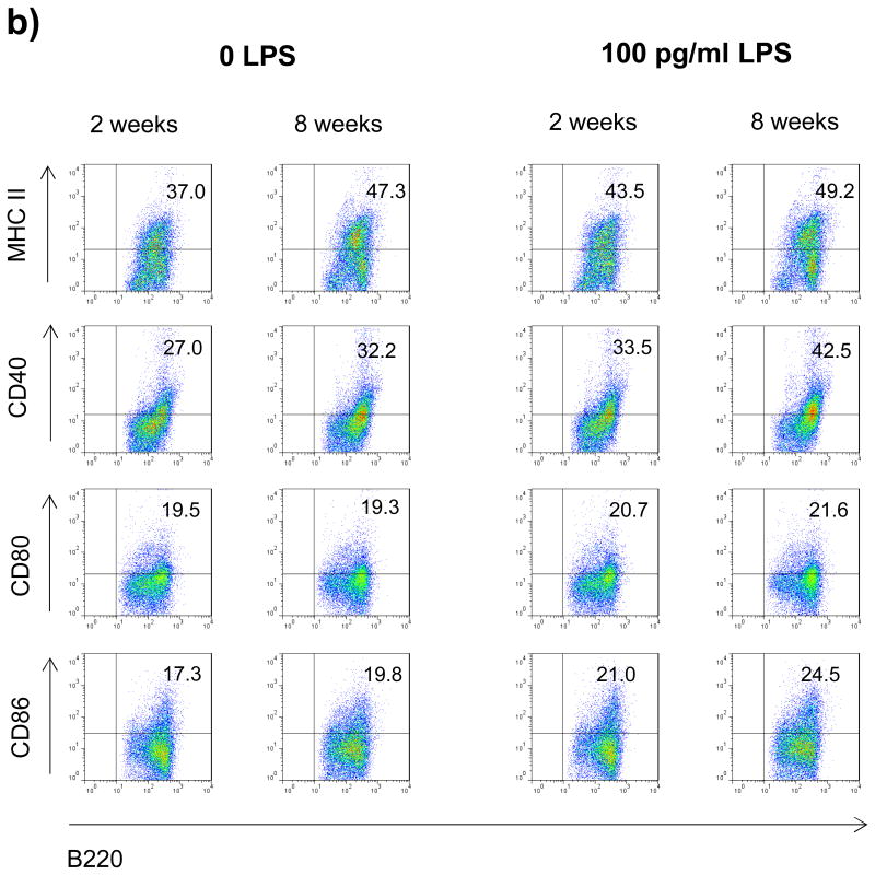
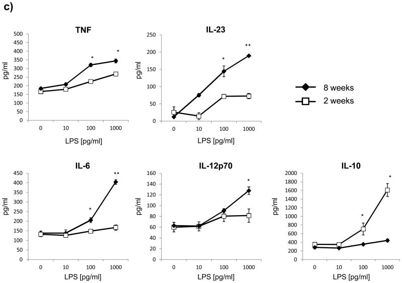
Splenocytes from 2 or 8 week old mice were cultured with 0 or 100 pg/ml LPS for 48h. Surface expression of MHC II and co-stimulatory molecules (CD40, CD80, CD86) by a) myeloid APC (CD11b+; mother gate viable leucocytes) and b) B cells (B220+; mother gate viable leucocytes) was evaluated by FACS staining. For a detailed description of the gating strategy see also suppl. Fig. 3. c) For evaluation of cytokine production splenocytes were stimulated with increasing concentrations of LPS and secretion of TNF, IL-6, IL-23, IL-10 and IL-12 was determined by ELISA after 72h. Data was obtained in triplicates and is presented as mean +/- SEM. Significance of differences of mean values was calculated using unpaired two-sided student's t-Test. * p ≤ 0.05; ** p ≤ 0.001. Shown is one representative out of two independent experiments.


