Abstract
Diseases affect coral species fitness and contribute significantly to the deterioration of coral reefs. The increase in frequency and severity of disease outbreaks has made evaluating and determining coral resistance a priority. Phylogenetic patterns in immunity and disease can provide important insight to how corals may respond to current and future environmental and/or biologically induced diseases. The purpose of this study was to determine if immunity, number of diseases and disease prevalence show a phylogenetic signal among Caribbean corals. We characterized the constitutive levels of six distinct innate immune traits in 14 Caribbean coral species and tested for the presence of a phylogenetic signal on each trait. Results indicate that constitutive levels of some individual immune related processes (i.e. melanin concentration, peroxidase and inhibition of bacterial growth), as well as their combination show a phylogenetic signal. Additionally, both the number of diseases affecting each species and disease prevalence (as measures of disease burden) show a significant phylogenetic signal. The phylogenetic signal of immune related processes, combined with estimates of species divergence times, indicates that among the studied species, those belonging to older lineages tend to resist/fight infections better than more recently diverged coral lineages. This result, combined with the increasing stressful conditions on corals in the Caribbean, suggest that future reefs in the region will likely be dominated by older lineages while modern species may face local population declines and/or geographic extinction.
Introduction
Immune defenses are critical for species success on ecological and evolutionary time scales [1]–[3]. As species diverge, new sets of genetic, biological and/or environmental conditions are encountered making it necessary for emerging species to trade off costs and benefits within and between traits [4], including those related to immunity [5]. Immunity plays an important role in the success of a given species and, in theory, evolves as species diverge [1], [6], likely conserving beneficial mechanisms from ancestral species [7]. Depending on selective pressures (e.g. resources and/or stressors), the immune system develops novel strategies and diversifies during speciation [6], [8], hence favoring individuals that survive pathogenic infections and other stressful events [7], [9]. In closely related species, the study of immunity in relation with phylogeny can provide insight into the selective forces at work and the way organisms and populations may respond to them [6], [10], [11].
The deterioration of coral reef ecosystems has been associated, among other factors, with changes in environmental conditions (e.g. increased water temperature and ocean acidification) and a significant increase in number of coral diseases and epizootics [12]–[16]. However, the evolutionary importance of immune traits in corals has not yet been evaluated. In other organisms such as fleas [17], termites [18] birds [19], [20], and vertebrates in general [21], immune traits are related to phylogeny. In corals, a recent taxonomic reorganization [22] facilitates assessment of trait variation and their relationship to life-history.
Scleractinia, the Cnidarian Order grouping all reef-building corals, is divided into two divergent groups termed Robust and Complex corals [23]–[26], each composed of several non-monophyletic families [27] with different evolutionary histories between the Atlantic and the Indo-Pacific regions [28]. Modern Caribbean scleractinians are grouped in at least six families, representing both Robust and Complex corals [27]. Recently, the Caribbean has become a disease ‘hot spot’ due to the high number of coral diseases, disease outbreaks and their widespread geographical range [16], [29], [30].
The innate immune system in corals is comprised of conserved components similar to those of other invertebrate [31], [32] and vertebrate species [33], [34] including the three general phases in the response to infection: recognition, signaling and effector responses [35]. While recognition receptors and several signaling pathways (e.g. toll and complement pathways) are activated upon pathogen recognition, many immune components, such as some effector mechanisms, show constitutive activity (i.e. non-pathogen induced or basal levels). Some of the better studied immune mechanisms in corals include the melanin synthesis cascade (e.g. prophenoloxidase and phenoloxidase) [31], [35], [36], antimicrobial compounds [31], [36]–[38], and antioxidants (e.g. superoxide dismutase, peroxidase and catalase) [31], [32], [35], [36]. Combined, these immune components provide corals with the capability to control the presence and combat proliferation of pathogens [31], repair tissue [39], and reduce levels of reactive oxygen species generated during infections and associated stress [40], [41]. Constitutive levels of prophenoloxidase, melanin [35] and antimicrobial activity [42] have been linked to disease resistance [32], [35], [43]–[45] suggesting an active investment in components of the immune system [10].
Investment in immunity has been related to different life history traits [1]. Since all biological traits, including those involved in immunity, tend to vary within and across populations and/or species during speciation, different evolutionary pressures (e.g. new pathogens or changes in climate and/or local environmental factors) can potentially result in distinct, species-specific, immune defenses. In corals, analyses of ecological strategies suggests related groups are affected similarly by environmental stressors and diseases [16], [46]–[48], but little is known about the role innate immunity plays in this pattern. Phylogenetic signals of biological traits are expected to be common across different groups of organisms [11], [49], but it is unknown what types of traits or what traits themselves will show a signal. Detecting a phylogenetic signal in coral immunity can provide insight into the evolution of the coral' immune system, and help explain the current pattern of disease resistance and its implications for the future of coral species and coral reefs, more so in light of global climate change and increased disease pressure [11], [50], [51].
In this study, we tested for the presence of a phylogenetic signal in immune traits and epizootics in Caribbean corals. We characterized the constitutive levels of six distinct immune traits in 14 of the most common and widely distributed coral species in the wider Caribbean. This represents ∼20% of the total diversity of scleractinian corals in the region [52]. We also compiled published disease data and determined the levels of immunocompetence of the studied species into two different metrics: Number of diseases (including both tissue loss diseases and growth anomalies) affecting each species, and mean prevalence, or the proportion of infected individuals in the population of a given species. These data sets were incorporated with the phylogeny of the host species (based on the 28S rDNA region) into three phylogenetic signal estimators (Bloomberg's K, Moran's I and Abouheif test).
The null hypothesis was that in scleractinian corals there is no relationship between phylogeny and levels of various enzymes involved in constitutive immunity and no correlation with disease parameters. Our results show co-variation between constitutive immunity and phylogeny in Caribbean corals, with species in older lineages (lineage defined as extant species and their ancestors) grouping together with lower number of disease and disease prevalence, than the modern lineages. Combined with other life history traits, these older lineages may be better suited to survive current and emerging diseases in the wider Caribbean.
Materials and Methods
Sample collection
For this study, 14 of the most common Caribbean scleractinian species comprising10 genera and 6 families [as defined by: 22, 27, 28, 53], were collected from several reefs (Media Luna - 17° 56.096 N; 67° 02.911 W, Turromote - 17° 56.097 N; 67° 01.130 W, Conserva - 17° 57.831 N; 67° 02.940 W, Pinnacles - 17° 55.963 N; 67° 00.714 W, Corral - 17° 56.986 N; 67° 00.504 W and Isla Cueva - 17° 57.599 N; 67° 04.827 W) off La Parguera, southwest coast of Puerto Rico (Table 1). To prevent seasonal or environmental effects, all samples were collected during the summer (northern hemisphere), the second week of August 2012. This collection represents approximately ∼20% of the total number of scleractinian coral species in the region [54]. Many of these species, i.e. Montastraea cavernosa, Orbicella spp. ( = Montastraea annularis complex), Diploria, Pseudodiploria, and Porites spp. are common and widely distributed through the region. Other groups, such as acroporids (A. palmata, A. cervicornis and A. prolifera) and pocilloporids (e.g. Madracis spp.) were not collected due to strict limits on sampling and manipulation.
Table 1. List of scleractinian coral species used to measure constitutive immunity and its variation across taxonomic levels.
| Family | Genus | Species | NCBI accession |
| Poritidae (III) | Porites | Porites astreoides | EU262830 |
| Poritidae (III) | Porites | Porites porites | EU262878 |
| Siderastreidae (IX) | Siderastrea | Siderastrea radians* | KJ946356 |
| Meandrinidae (XII) | Dendrogyra | Dendrogyra cylindrus | EU262819 |
| Meandrinidae (XII) | Meandrina | Meandrina jacksoni* | KJ946355 |
| Meandrinidae (XII) | Meandrina | Meandrina meandrites | EU262815 |
| Montastraeidae (XVI) | Montastraea | Montastraea cavernosa | EU262810 |
| Merulinidae (XVII) | Orbicella | Orbicella annularis | HQ203479 |
| Merulinidae (XVII) | Orbicella | Orbicella franksi | EU262849 |
| Merulinidae (XVII) | Orbicella | Orbicella faveolata | EU262781 |
| Mussidae (XXI) | Diploria | Diploria labyrinthiformis | EU262772 |
| Mussidae (XXI) | Pseudodiploria | Pseudodiploria strigosa* | KJ946354 |
| Mussidae (XXI) | Mussa | Mussa angulosa | EU262869 |
| Mussidae (XXI) | Mycetophyllia | Mycetophyllia aliciae | EU262809 |
Underlined groups within the family and genus categories represent the sequence used for that group in the phylogenetic signal assessments. Accession numbers correspond to the sequence of the 28S rDNA region used for each species. (* = Species sequenced in this project).
Small fragments from a total of 140 apparently healthy (i.e. with no signs of disease or bleaching) colonies (10 per species) were sampled. All samples were collected under the specification of research collection permits to the Department of Marine Science University of Puerto Rico – Mayagüez (UPRM), issued by the Department of Natural Resources of Puerto Rico. A fragment of approximately 5 cm2 was carefully removed from the top of each massive/crustose colony with a hammer and a chisel. Small branches were broken from branching colonies. For the “free-living” Siderastrea radians, rolling stones larger than 4 cm in diameter were collected in shallow sea-grass beds next to the reefs. All samples were stored in individually labeled sterile Whirl-pack bags (Fisher Scientific, Waltham, MA), transported in seawater to the laboratory and flash-frozen in liquid nitrogen. Frozen samples were stored at −80°C, shipped to the University of Texas at Arlington (UTA) in dry ice and kept at −80°C until further analyses.
DNA extractions and PCR amplifications
The NCBI database has a significant number of sequences from most of the corals used in this project. The 28S rDNA region has been sequenced (as of September 2013) for 11 of the 14 species in this study (Table 1), and phylogenetic reconstructions showing similar topologies to other molecular markers, and divergence time estimation using fossils (Caryophyllia spp., Flabellum spp. and Dendrophyllidae), were available. Sequences for three species (Meandrina jacksoni GenBank KJ946355, Pseudodiploria strigosa KJ946354 and Siderastrea radians KJ946356) were generated in this project after extraction of DNA using a modified protocol from LaJeunesse et al. [55]. A small fragment (∼3 mg) of skeleton and tissue was mixed with (of glass beads (∼200 µl, 1 mm, Ceroglass, Columbia, TN) and 600 µl of a cell lysis solution (0.2 M Tris, 2 mM EDTA, 0.7% SDS, pH 7.6) and shaken on a BioSpec (Bartlesville, OK) beadbeater for 100 seconds. Proteinase K (3 µl −20 mg/ml) was added and incubated at 65°C for 1 hour. The incubation was followed by protein precipitation with ammonium acetate (250 µl −9 M) and freezing at −20°C. The frozen extract was centrifuged (10,000 G for 15 minutes) and the supernatant removed, mixed with 600 µl of isopropanol (100%) and centrifuged (10,000 G for 5 minutes). The DNA pellet was washed with 70% ethanol, air dried, and resuspended in 75 µl of distilled water and stored at −20°C.
The 28S rDNA region was amplified using the 28SROM.IFw (5′-GGCGACCCGCTGAATTCAAGCATAT-3′) and 28SDES.VRv 5′-GGTCTTTCGCCCCTATACTC-3′) primers [56]. Reactions were performed using Perfect Taq Plus DNA Polymerase (5-Prime, Gaithersburg, MD) following the manufacturer recommended reaction composition on 2 µl of 1∶40 dilutions of the extracted DNA (final reaction volume 25 µl). Amplifications consisted of 35 cycles of 95°C, 52°C and 72°C steps, each for 30 seconds. Amplified products were cleaned with ExoSap (Affymetrix, Santa Clara, CA) and sequenced with the forward primer using Big Dye 3.1 terminator mix (Applied Biosystems/Life Technologies, Grand Island, NY) on an ABI Hitachi 3730XL genetic analyzer at UTA' Genomics Core Facility. DNA sequence chromatograms were reviewed and edited using Geneious Pro 5.0 [57]. The resulting sequences were combined with those obtained from the NCBI data base (657 to 685 bp) and alignments were performed on ClustalW using a gap-opening penalty of 15 and a extension penalty of 6 [58]. Phylogenies were constructed on MrBayes [59], using a general time-reversible model with gamma distributed rate heterogeneity (GTR+G) as substitution model, a chain length of 1,100,000 and 100,000 burn-in (phylogenies can be found in Text S1).
In order to determine the approximate age of the studied lineages, that is the extant species and their ancestors, we calculated the divergence times on the 28S rDNA based phylogeny (Text S2). Divergence times were determined with BEAST 1.7.5 [60], using a relaxed-clock uncorrelated lognormal allowing for nucleotide substitutions rates to vary between lineages [26]. The tree prior used a Yule process and the model of substitution was set to gamma distributed rate heterogeneity as suggested by jModeltest 2.1.3 [61], with invariant sites (GTR+G+I). Node ages and posterior probabilities were estimated on a run with 10,000,000 generations and saving the topologies and parameters every 1,000 generations. In order to estimate the early divergence of the studied groups, calibrations were done using Dendrophyllidae (∼127 Mya; including sequences of Tubastrea coccinea, Cladopsamia gracilis, Leptosammia pruvoti, Endopachys grati, Enollopsamia rostrata and Balanophyllia spp.), Caryophyllia spp. (∼160 Mya) and Flabellum spp. (∼77.5 Mya) as suggested by Stolarski et al. [26]. Phylogenetic reconstruction within BEAST was performed using Mean heights and node heights, a prior probability of 0.1 and 1,000,000 burn-in.
Protein extractions and immune assays
Protein extractions and enzymatic assays followed protocols previously used to study coral immunity [31], [32], [35], [36], [43]. Coral tissue was airbrushed from the skeleton using a Paasche single action artistic airbrush (Paasche Airbrush Company, Chicago, IL) with minimal amounts (5–6 ml) of Tris buffer (100 mM Tris, pH 7.8+0.5 mM dithiothreitol). To break open cells and extract proteins, tissue slurries were homogenized using a tissue homogenizer (Powergen 125, Fisher Scientific, Waltham, MA) for 1 minute on ice. One ml of the tissue slurry was added to pre-weighted 1.5 ml microfuge tubes for melanin concentration estimates. All homogenates were centrifuged at 90×G for 10 minutes and the supernatant was recovered. Protein concentrations were estimated using the RED660 protein assay (G Biosciences, Saint Louis, MO) and standardized to a standard curve of bovine serum albumin.
We performed assays for six immune traits: prophenoloxidase, melanin concentration, superoxide dismutase, peroxidase, catalase activity and inhibition of bacterial growth. Prophenoloxidase was tested on 20 µl of the extract, mixed with 20 µl of sodium phosphate buffer (50 mM, pH 7.0) and 25 µl of Trypsin (0.1 mg/ml). The reaction was initiated by adding 30 µl of dopamine (10 mM) as a substrate. Change in absorbance was measured every 30 seconds at 490 nm for 15 minutes and activity calculated during the linear range of the curve (1–5 minutes).
Melanin concentration was assessed on the melanin-reserved portion of initial tissue slurry after freeze-drying (VirTis BTK, SP Scientific, Warminster, PA) for 24 hours. The resulting dried tissue was weighed and the melanin extracted with 400 µl NaOH (10 M). Extraction was done at room temperature for 48 hours at which time the tissue particles were centrifuged (90×G) for 10 minutes. 60 µl of the supernatant were used to determine the absorbance at 410 nm. Resulting values were standardized to a dose-response curve of commercial melanin (Sigma-Aldrich, Saint Louis, MO).
Superoxide dismutase activity was determined with the SOD Determination kit (#19160, Sigma-Aldrich, Saint Louis, MO) following manufacturer's instructions. Absorbance at 450 nm was measure in wells containing coral protein extracts and superoxide dismutase controls and compared to untreated samples. The inhibition was normalized by mg protein and presented as superoxide dismutase activity units per mg protein.
Peroxidase activity was assessed on 10 µl of extract with 40 µl phosphate buffer (0.01 mM, pH 6.0) and 25 µl Guaiacol (25 mM). Activity was monitored for 15 minutes, recording the absorbance at 470 nm every 30 seconds. Peroxidase is presented as change in absorbance per mg protein per minute.
Catalase was measured as the change in hydrogen peroxide concentration after mixing 5 µl of the protein extract with 45 µl of sodium phosphate buffer (50 mM, pH 7.0) and 75 µl of 25 mM H2O2. Samples were loaded on UV transparent plates (Grainer Bio-one, Monroe, NC) and read at 240 nm every 30 seconds for 15 minutes. Catalase activity was estimated as change in hydrogen peroxide concentration per mg of protein during the first minutes of the reaction.
The percent inhibition of bacterial growth was assessed against Vibrio alginolyticus (Strain provided by K. Ritchie, Mote Marine Laboratory, GenBank # X744690). This particular bacterial strain was isolated from Orbicella faveolata, and has been implicated in Yellow Band Disease [62], [63], and thus offers a good estimate of antibiotic activity in a broad range of corals. Bacteria were grown in salt amended (2.5% NaCl) Luria Broth (EMD Chemicals, Gibbson, NJ) for 24 hours prior to use in the assay. The resulting bacteria culture was diluted to a final optical density of 0.2 at 600 nm. 140 µl of the culture suspension was added to each well along with 60 µl of the coral extract. To detect possible effects of the media and the airbrushing buffer, controls with 60 µl of Tris Buffer (100 mM Tris, pH 7.8+0.5 mM dithiothreitol) were included on each experimental plate. Plates were incubated in the spectrophotometer for 6 hours at 29°C, determining the absorbance at 600 nm every 10 minutes. The change in absorbance during the logarithmic phase were used to determine the growth rate of each samples and the proportion of this rate to that of the bacterial control provided the percent inhibition for each samples.
These immune assays are ideal for comparative immunity studies because they are not species-specific [10]. All assays were conducted on a Synergy 2 Microplate Reader (Biotek Instruments, Winooski, VT) and standardized to mg of protein when applicable. Constitutive levels of all six immune traits were compared between older and modern coral lineages (groups described in the results section). Comparisons were assessed with a t-test assuming unequal variances across traits.
Data analysis and phylogenetic signal estimations
In order to test the hypothesis that closely related taxa have similar activity of constitutive immune components, the data was partitioned in three groups corresponding to each of the clades of interest: family, genus and species (Table S1). Results from the immune assays were averaged for each taxon on each level. Additionally, to obtain an integrated measure of immunity the first component scores from a principal component analysis (PC1) with all individual immune measures was used as an additional category. Principal components reduce dimensions and convert multiple variables into composite indicators [64], improving the analysis of immune capacity in relation to other biological/environmental parameters [65]. The analyses were done in JMP 10.0.0 (SAS Institute, Cary, NC).
In order to detect a phylogenetic signal, the immune data sets were compared against family, genera and species phylogenetic reconstructions (Text S1). The genus and family phylogenies were built with subsets of selected sequences from each group (underlined categories in Table 1). For example, Pseudodiplora strigosa 28S sequence was used as the representative for the Pseudodiplora genus.
The possibility of phylogenetic signals in coral constitutive immune levels was tested with Bloomberg's K [49] and Moran's I measures as described by Gittleman and Kot [66] and by Abouheif test [67]. K assumes the data follows a Brownian motion model (BM) and compares the observed phylogenetic signal with that of the trait under the BM model. Higher K values for a particular trait represent a stronger phylogenetic signal, and zero values indicate no effect of phylogeny [17], [49]. Moran's I (I) on the other hand, is a model-independent measure of autocorrelation in which the relation between the variation in the trait and the phylogenetic distance is established. In this method, the data is divided into the phylogenetic component and the trait component and correlarograms are built to determine the effect of ranks and distances [66]. Lastly, the Abouheif [67] test (A), modifies Moran's I to successfully detect phylogenetic signal of different traits on phylogenies with both low and high number branches. All phylogenetic signal tests were performed on R using geiger, carper, picante, adephylo and phylobase packages.
A Spearman's rank order correlation index was used to determine correlations between each immune measure and estimates of number of diseases and prevalence (proportion of infected individuals in the population of a given species at a given time) for each taxonomic level (i.e. species, genus and family). Disease parameters (number of diseases and prevalence) were compiled from literature reporting epizootic events in the Caribbean from 1997 to 2005. Only manuscripts with species level resolution, and infected or diseased colonies specified as a subset of the total population surveyed were used so that number of diseases and prevalence estimates could be normalized across reports. Prevalence was estimated from the reports by combining data from all surveyed diseases (e.g. Black Band, White Band, White Plague, Yellow Band, Dark Spots, and growth anomalies) for each species. A total of 12 reports with appropriate information were found from different locations in the Caribbean, including Bermuda, Florida, Bahamas, Puerto Rico, Saint Croix, Bonaire, Yucatan-Mexico, Colombia and Venezuela (Table 2) [29], [30], [68]–[77]. The geographic coverage and extent of the disease surveys in these reports provided a very robust data set for both number of diseases and disease prevalence, thus truly representing the disease dynamics across the region. Phylogenetic signal in the number of diseases and prevalence was estimated as described above.
Table 2. List of references, reports and reviews of coral disease parameters from various locations in the Caribbean used to obtain data for the 14 species used in the phylogenetic signal estimates.
| Reference | Location | Survey year(s) | Data used |
| Sutherland et al., 2004 | Caribbean Wide | Review | Number of diseases |
| Weil, 2004 | Caribbean Wide | Review of 1999–2002 | Number of diseases and prevalence |
| Kaczmarski et al., 2005 | Saint Croix | 2001 | Prevalence |
| Voss and Richardson, 2006 | Lee Stocking Island - Bahamas | 2002, 2003 | Prevalence |
| Santavi et al., 2001 | Florida | 1997, 1998 | Number of diseases |
| Ward et al., 2006 | Yucatan Peninsula - Mexico | 2004 | Prevalence |
| Garzón-Ferreira et al., 2001 | Colombia | 1998 | Number of diseases |
| Navas-Camacho et al., 2010 | Colombia | 1998–2004 | Number of diseases |
| Gil-Agudelo et al., 2010 | Colombia | 1998–2005 | Number of diseases |
| García et al., 2003 | Los Roques - Venezuela | 1999 | Number of diseases and prevalence |
| Croquer et al., 2003 | Los Roques - Venezuela | 2000 | Number of diseases and prevalence |
| Croquer et al., 2005 | Los Roques - Venezuela | 2000, 2001 | Prevalence |
Data used included the number of diseases affecting each species and average disease prevalence (proportion of infected individuals on a population of a given species) for all diseases affecting each species.
Results
Phylogenetic reconstruction and time of divergence
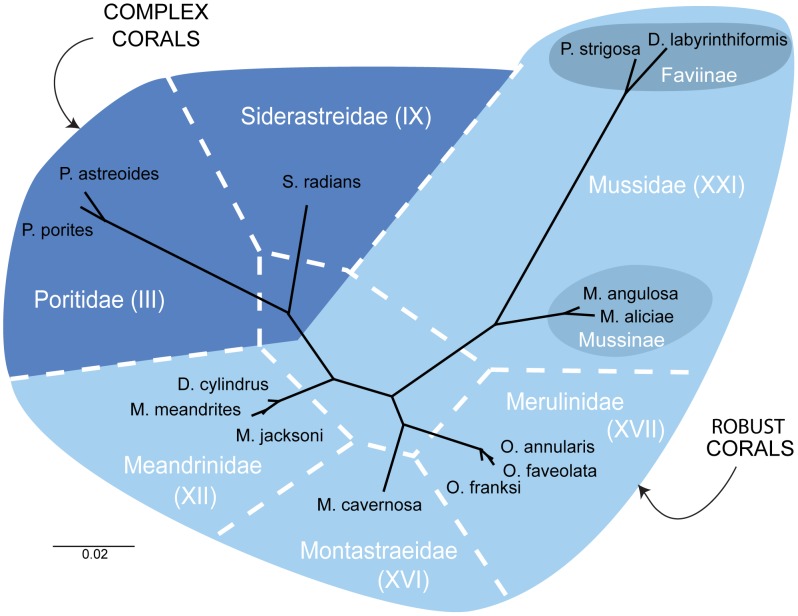
Sequences of the 28S rDNA region for three corals were generated and in each case these newly sequenced species clustered according to current taxonomy; Meandrina jacksoni with its sister species M. meandrites, Pseudodiploria strigosa with Diploria labyrinthiformis among the robust corals and Siderastrea radians with Porites spp. in the complex corals (Figure 1). The phylogenetic hypothesis obtained with the 28S rDNA region, using all 14 species, resolved the same groups delineated with other molecular markers and in other geographic locations [27], [53], [78].
Figure 1. Un-rooted Bayesian phylogenetic reconstruction (dark line) using the 28S rDNA (657–685 bp) region from 14 species of Caribbean corals used in this study.
The dotted lines represent the family groupings as recently proposed by Budd et al. [22]. All nodes in this tree have support (posterior probability) values higher than 0.80.
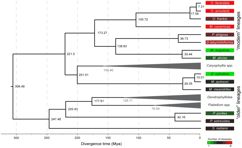
Divergence times of the included lineages (i.e. extant species and ancestors) derived from the molecular clock show that Siderastrea radians (∼247 Mya) and Porites spp. (∼220 Mya) diverged early during the Triassic and appear as ancestral lineages among the Complex corals. Among the robust corals, the family Meandrinidae also showed early divergence (∼201 Mya), followed by a recent radiation into the current species (∼30–42 Mya; Figure 2). The other Complex lineages evolved within the last ∼100 to 150 Mya (Figure 2). Faviinae (D. labyrinthiformis and P. strigosa) and Mussinae (Mycetophyllia aliciae and Mussa angulosa) diverged ∼138 Mya, with current species diverging as recently as 40 Mya. The genera Orbicella and Montastraea originated ∼105 Mya, with Orbicella radiating between ∼17 and ∼7 Mya (Figures 1 and 2). This allowed the sampled lineages to be grouped into older (S. radians, P. porites, P. astreoides, D. cylindrus, M. meandrites and M. jacksoni) and modern (M. aliciae, M. angulosa, D. labyrinthiformis, P. strigosa, M. cavernosa, O. faveolata, O. annularis and O. franksi) lineages.
Figure 2. Phylogenetic reconstruction with the 28S rDNA region used to estimate the divergence time (dark values in the nodes) of the evolutionary lineages of 14 Caribbean coral species.
Time of divergence was estimated with BEAST following the method by Stolarski et al. [26]. The molecular clock was calibrated with specimens from the family Dendrophyllidae at 127±3.5 Mya, and the genera Caryophyllia at 160±3.5 Mya and Flabellum at 77.5±3.5 Mya, represented by gray triangles and values. Number of diseases affecting each species is shown in the colored squares. Disease data was compiled from the literature (Table 2) and corresponds to surveys between 1997 and 2005 throughout the Caribbean.
Phylogenetic signal in coral immunity and disease
Results from the three phylogenetic signal measures were similar across taxonomic levels (i.e. species, genus and family). These analyses revealed different patterns of phylogenetic signal for each independent immune measure (Table 3). Significant correlations between phylogeny and immune traits were found in melanin concentration at the species level (Moran's I - I sp = 0.105, p = 0.036), peroxidase activity at the genus (I genus = 0.451, p = 0.006; A genus = 0.524, p = 0.005) and species levels (Bloomberg's K - Ksp = 1.269, p = 0.001; I sp = 0.488, p = 0.002; A sp = 0.583, p = 0.002) and percent inhibition of bacterial growth at the family level (Kfamily = 1.637, p = 0.010, I family = 0.082, p = 0.048). Variation in prophenoloxidase, superoxide dismutase and catalase activities did not show a significant phylogenetic signal at any level (Table 3).
Table 3. Bloomberg's K, Moran's I and Abouheif I test (A) values, testing for phylogenetic signal in constitutive immunity (six individual measures and the integrated measure - PC1) and number of diseases affecting each species and prevalence (proportion of individuals infected in the population of a given species) at three taxonomic levels (family, genus and species) within 14 scleractinian coral species from the Caribbean Sea (bolded values show significant phylogenetic signal).
| Individual immune measures | Integrated Measure | Disease Parameters | |||||||
| Melanin Synthesis Pathway | Antioxidants | Antimicrobial Activity | |||||||
| Prophenol oxidase | Melanin | Superoxide dismutase | Peroxidase | Catalase | % Inhibition of bacterial growth | PC1 | Number of diseases | Prevalence | |
| Family | |||||||||
| K | 0.969 | 1.328 | 1.106 | 1.474 | 0.932 | 1.637 | 1.080 | 1.262 | 0.785 |
| I | −0.018 | −0.139 | −0.171 | 0.218 | −0.236 | 0.082 | −0.130 | 0.153 | 0.106 |
| A | 0.116 | 0.382 | 0.101 | 0.326 | 0.030 | 0.266 | 0.122 | 0.248 | 0.182 |
| Genus | |||||||||
| K | 0.217 | 0.874 | 0.890 | 0.903 | 0.407 | 0.848 | 0.962 | 0.923 | 0.460 |
| I | −0.067 | −0.156 | 0.067 | 0.451 | −0.032 | 0.187 | 0.219 | 0.226 | −0.036 |
| A | 0.039 | −0.062 | 0.247 | 0.524 | 0.008 | 0.252 | 0.325 | 0.358 | 0.011 |
| Species | |||||||||
| K | 0.252 | 0.168 | 0.534 | 1.269 | 0.329 | 0.321 | 0.504 | 0.747 | 1.102 |
| I | −0.110 | 0.105 | 0.188 | 0.488 | −0.020 | 0.182 | 0.251 | 0.292 | 0.370 |
| A | −0.041 | 0.114 | 0.213 | 0.583 | 0.005 | 0.202 | 0.271 | 0.351 | 0.544 |
Principal Component Analysis revealed that Principal Component 1 (PC1) explained 33.5% of the variation, Principal Component 2 (PC2) 25.1% and Principal Component 3 (PC3) 23.3%. PC1 values were similar between closely related species showing a significant phylogenetic signal at the genus (A genus = 0.325, p = 0.037) and species (I sp = 0.251, p = 0.036; A sp = 0.271, p = 0.049; Table 3) levels. PC2 and PC3 did not show a significant phylogenetic signal.
Closely related species have similar number of diseases and disease prevalence (proportion of individuals infected in the population of a given species). The number of diseases showed significant phylogenetic signal at the genus (A genus = 0.358, p = 0.037) and species (K sp = 0.747, p = 0.015, I sp = 0.292, p = 0.026; A sp = 0.351, p = 0.019) levels. Variation in disease prevalence was significant at the family (I family = 0.106, p = 0.012) and species (Ksp = 1.102, p = 0.009; I sp = 0.370, p = 0.004; A sp = 0.544, p = 0.007) levels (Table 3).
Correlations between disease parameters and immune measures
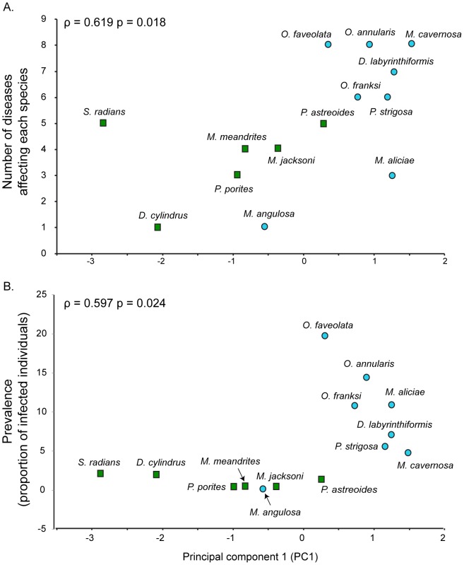
Several of the individual immune traits were significantly correlated to number of diseases and prevalence. Peroxidase (ρ = 0.971, p = 0.001) at the family level, prophenoloxidase (ρ = 0.667, p = 0.035), superoxide dismutase (ρ = 0.636, p = 0.048) at the genus level and prophenoloxidase (ρ = 0.564, p = 0.036) at the species level showed significant correlations with number of diseases. Only peroxidase (ρ = 0.971, p = 0.001) at the family level was correlated with disease prevalence (Table 4). The integrated measure of immunity (represented by PC1) was positively and significantly correlated with number of diseases (ρ = 0.619, p = 0.018) and prevalence (ρ = 0.597, p = 0.024; Figure 3) at the species level. Number of diseases increases as divergence time of the lineages decrease.
Table 4. Spearman's rank order correlations (ρ) between constitutive levels of six immune traits and number of diseases affecting each species and average prevalence (proportion of infected individuals on a population of a given species) of all diseases affecting each species, at three taxonomic levels (family, genus and species) among 14 scleractinian coral species from the Caribbean (significant correlations are bolded).
| Individual immune traits | Integrated measure | ||||||
| Melanin Synthesis Pathway | Antioxidants | Antimicrobial Activity | |||||
| Prophenol oxidase | Melanin | Superoxide dismutase | Peroxidase | Catalase | % Inhibition of bacterial growth | PC1 | |
| Number of diseases | |||||||
| Family | 0.618 | 0.000 | 0.530 | 0.971 | 0.088 | −0.795 | 0.706 |
| Genus | 0.667 | 0.073 | 0.636 | 0.422 | 0.428 | −0.294 | 0.593 |
| Species | 0.564 | −0.107 | 0.517 | 0.531 | 0.264 | −0.340 | 0.619 |
| Disease prevalence | |||||||
| Family | 0.618 | 0.000 | 0.530 | 0.971 | 0.088 | −0.795 | 0.706 |
| Genus | 0.309 | 0.030 | 0.394 | 0.261 | 0.515 | −0.491 | 0.588 |
| Species | 0.265 | −0.128 | 0.358 | 0.484 | 0.230 | −0.475 | 0.597 |
Figure 3. Correlation between (A) number of diseases (number of diseases affecting each coral species) and (B) prevalence (proportion of individuals infected in the population of a given species) with constitutive levels of immunity as measured with the Principal Component 1 (PC1) obtained from Principal Component Analysis of six immune measures (prophenoloxidase, melanin concentration, superoxide dismutase, peroxidase, catalase and inhibition of bacterial growth).
The correlations were determined with the Spearman's rank order index (ρ) for both disease susceptibility and prevalence. Green squares represent older, and blue circles modern lineages.
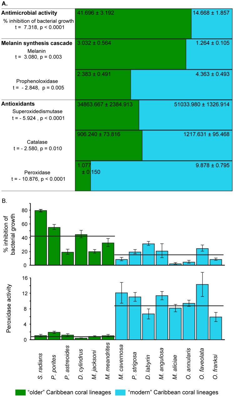
Older lineages such as Porites spp. (known to be affected by at least 5 diseases), Siderastrea radians (5 diseases) and the Meandrinidae species (4 diseases) are affected by fewer diseases than modern groups such as Diploria (7 diseases), Pseudodiploria (6 diseases), Montastraea (8 diseases) and Orbicella (8 diseases; Figure 2) [29], [30], [68]–[75], [79]. Species from the subfamily Mussinae (Mycetophyllia aliciae and Mussa angulosa) were the exception with an average divergence time of ∼33 Mya and a low susceptibility (3 and 1 diseases respectively). The fact that Mycetophyllia and Mussa form a well-supported monophyletic group [22], [27], [78] that according to our molecular clock diverged ∼138 Mya (Figure 2), may explain this inconsistency. Pairwise (t-tests) comparisons showed all six immune traits levels are significantly different (p<0.050) between older and modern lineages (Figure 4).
Figure 4. Proportion of the mean (± standard error) values of constitutive levels of six immune traits (percent inhibition of bacterial growth, melanin concentration, prophenoloxidase, superoxide dismutase, peroxidase and catalase activity) for older and modern Caribbean coral lineages (A).
Pairwise comparisons for each trait using t-tests assuming unequal variances revealed significant differences between older and modern lineages for all traits. Older lineages showed higher mean values of melanin and percent inhibition of bacterial growth and lower values in the other traits compared to the modern lineages. Percent inhibition of bacterial growth and peroxidase activity (mean ± standard error of change in absorbance 490 nm per mg protein per minute) for 14 Caribbean coral species (B). Black lines represent the average immune assay values for each coral lineage.
Discussion
Immune defenses are key to the evolutionary success of a species [3], but does the immune system retain ancestral characteristics, develop new or retain both ancestral and new defense strategies during speciation? [7] Answering this question in scleractinians is important since it can offer insight into the evolution of immunity and relevance to disease resistance and the future of corals in light of novel pathogens and climate change [50]. Results of this study indicate that the coral immune system, at least in Caribbean corals, has been molded by evolutionary history, with past selective pressures (biotic and abiotic) likely improving the innate immune system to respond more efficiently to new diseases [80]. Extant species, belonging to groups of species that have survived through more of these pressures, i.e. older coral lineages (Porites spp. ∼220 Mya, Siderastrea radians ∼247 Mya and Meandrinidae spp. ∼201 Mya), seem to be evolutionarily better equipped to cope/survive current conditions than more recent or modern lineages (Orbicella spp. = Montastraea annularis complex, ∼105 Mya).
Constitutive immunity, disease patterns and phylogenetic analyses of the 14 coral species in this study show some interesting patterns. Species from lineages that diverged more than 200 Mya are affected by fewer diseases, show lower disease prevalence, and have higher levels of some constitutive immune defenses. Some of these species, for example, P. astreoides and S. radians have ecological traits consistent with a resistant coral. Both are able to maintain normal physiological functions under stress [81], are brooders and exhibit a weedy-like dispersal and recruitment strategies [82]. Together with their high constitutive immune levels, these traits make for resistant and successful species [83] increasing survivorship and probably, species fitness.
Some of the modern lineages comprise another distinct group. Species in this group are affected by up to 8 different diseases, show high disease prevalence levels and have lower constitutive levels of some immune defenses. Orbicella faveolata, O. annularis, Diploria labyrinthiformis, and Pseudodiploria strigosa are among the species most heavily affected by disease in the Caribbean [16], [29], [30], leading to significant population losses in recent years.
This study did not include the Caribbean Acroporids (Acropora palmata and A. cervicornis). Acroporids in general are thought to have very low levels of immunity [35] and the Caribbean species were nearly extirpated over their geographic distribution by White Band Disease [84], [85]. High rates of clonal reproduction (i.e. low genetic variability) can result in higher population susceptibility to environmental and/or biological stressors [86]. Their clonal population structure [87] and life history traits (e.g. broadcasters with low recruitment success, fast growing branching morphology and high fragmentation rates, etc.) have rendered Caribbean acroporid populations highly susceptible to disease and contributed to their significant losses over their entire geographic distribution.
Overall, there was a relationship between constitutive levels of immune measures and diseases data that partitioned between old and modern lineages. Since the immune system is integrated, assessing several measures of immunity is ideal to gain a better understanding of both the potential to prevent and the capacity to fight infections [88], [89]. This study did not include induced immune responses or resistance to pathogens in experimental settings. However, it appears our multivariate immune measures are good indicators of disease susceptibility across these 14 species. Similar patterns to those detected here have been reported for Indo-Pacific corals, where specific markers of the melanin synthesis cascade were related to disease prevalence [35]. Our study integrates the melanin synthesis cascade, several antioxidants and general antimicrobial activity.
Among the specific immune traits, inhibition of bacterial growth and melanin concentration were higher in the older coral lineages, suggesting that these two mechanisms (among others not described here) may be important in conferring resistance. Prophenoloxidase and all the antioxidants were higher in the modern lineages. There is an inverse relationship between the melanin product and proteins in its biosynthetic cascade. Prophenoloxidase activity is likely to increase upon microbial stimulation, but corals with higher melanin concentrations may be able to prevent infection by deploying a melanin barrier before the need to engage into additional, more costly responses [90].
All antioxidants were more active in the modern lineages than in the older coral lineages. Antioxidants are an integral part of the stress response to both abiotic and biotic stressors. All modern lineages in this study are known to be susceptible to both elevated temperatures and to disease [31], [36], [43]. Increasing temperatures and/or pathogen load may keep these corals under a constant state of stress, thus elevating the levels of antioxidants at a cost to other defense pathways [32]. In addition to continuous stress, the algal symbiont type in each coral species may also play a part in oxidative stress that is reflected in the holobiont [91].
Variation in constitutive immunity among corals can also be the result of environmental and spatial gradients on a given reef. While our data are from one region and one time point, we fell they are a start to developing hypotheses to why older corals may be more resistant to diseases. In fact, we attempted to reduce the effects of the environment by collecting during the same season (within a week), and either sampling the same species from different reefs and different species from the same reef and from the same location of the colony. The exception was Siderastrea radians, which was collected in sea grass beds outside the reefs. However, S. radians immune measures clustered with those from both Porites porites and P. astreoides, supporting our hypothesis and minimizing the influence of the environment.
Associated organisms, Symbiodinium spp. and bacterial symbionts or communities may exert an important role in the immune defense of the coral host. Our protein extraction is enriched in host proteins but may include a minimal amount of Symbiodinium protein. The corals in this study associate with a variety of Symbiodinium species/strains (clades A, B, C, D). At least seven of the 14 coral species (e.g. Pseudodiploria strigosa, Dendrogyra cylindrus, Meandrina meandrites, M. jacksoni, Orbicella faveolata, O. franksi and O. annularis) are known to associate with Symbiodinium B1 [92]. The immune measures of these corals reflect the phylogeny of the coral host more than the presence of a particular symbiont.
Caribbean reefs are not likely to disappear, but rather change in community composition and structure [50], [93]. Coral species with advantageous life history traits will be more likely to dominate [94] with the immune capacity of individuals and/or populations serving as a critical piece of the puzzle. While our data are limited in the number of species and to the Caribbean region, the evidence presented here indicates that phylogenetically close scleractinians have comparable levels of constitutive immunity.
Our results support the ongoing observations that disease resistance varies considerably among coral taxa and provides a possible mechanistic basis for this variability. Caribbean species from older lineages seem better adapted to resist infections with high levels of melanin and antimicrobial activity and, as a result, may have experienced significantly lower mortality from disease outbreaks in the recent past [95]–[97]. The life history traits of many of these older lineages seem to make them well suited for survival under the current changing conditions. Correspondingly, older lineages of mammals appear less extinction-prone than previously predicted [98]. Recently diverged, modern Caribbean coral lineages on the other hand, have been severely affected by diseases and/or thermal stress with significant tissue loss and colony mortalities at local and regional geographic scales and some of these taxa could face local extinction in many areas [99].
Supporting Information
Mean values (± standard error) of six immune traits (prophenoloxidase, melanin, superoxide dismutase, peroxidase, catalase and percent inhibition of bacterial growth), the integrated immune measure (PC1) and disease parameters (number of diseases and prevalence) used in the phylogenetic signal analyses on 14 Caribbean corals at three taxonomic levels (species, genus and family).
(XLSX)
Phylogenetic reconstructions based on the 28S rDNA region of 14 Caribbean corals at three different taxonomic levels (species, genus and family). These trees were used as input in the phylogenetic signal analyses.
(NEWICK)
Sequence alignment of the 28S rDNA region used to determine the divergence times of 14 Caribbean corals. Dendrophyllidae (∼127 Mya; including sequences of Tubastrea coccinea, Cladopsamia gracilis, Leptosammia pruvoti, Endopachys grati, Enollopsamia rostrata and Balanophyllia spp.), Caryophyllia spp. (∼160 Mya) and Flabellum spp. (∼77.5 Mya) were used as calibration point in the molecular clock.
(NEX)
Acknowledgments
The authors would like to acknowledge the Department of Marine Sciences, University of Puerto Rico Mayagüez (UPRM) for partial funding for boat and diving, logistical support and laboratory space. The authors would also like to thank Derek Soto, Duane Sanabria and Luis Rodriguez (UPRM) for their help with collection and sample manipulations, Whitney Mann and the staff of the Genetics Core Facility (UTA) support in the field and laboratory work respectively. Comments by Matthew Fujita (UTA) and Jean-Francois Flot (MPI-DS) and two anonymous reviewers helped improve this manuscript. The analyses were run on a server provided by the UTA Office of Information Technology. Samples were collected under the specification of research collection permits to the Department of Marine Science UPRM.
Data Availability
The authors confirm that all data underlying the findings are fully available without restriction. All relevant data are within the paper and its Supporting Information files.
Funding Statement
Funding was provided by NSF grant IOS # 1017458 to L.D.M., NSF grant IOS # 1017510 and OCE -1105143 to E.W., and NSF grant OCE-PRF # 1225163 to J.H.P.C. The funders had no role in study design, data collection and analysis, decision to publish, or preparation of the manuscript.
References
- 1. Previtali MA, Ostfeld RS, Keesing F, Jolles AE, Hanselmann R, et al. (2012) Relationship between pace of life and immune responses in wild rodents. Mol Ecol Res 121: 1483–1492. [Google Scholar]
- 2. Schulenburg H, Kurtz J, Moret Y, Siva-Jothy MT (2009) Introduction. Ecological immunology. Philosophical Transactions of the Royal Society of London, Series B: Biological Sciences 364: 3–14. [DOI] [PMC free article] [PubMed] [Google Scholar]
- 3. Satake H, Sekiguchi T (2012) Toll-Like receptors of deuterostome invertebrates. Front Immunology 3: 1–7. [DOI] [PMC free article] [PubMed] [Google Scholar]
- 4. Norris K, Evans MR (2000) Ecological immunology: life history trade-offs and immune defense in birds. Behav Ecol 11: 19–26. [Google Scholar]
- 5. Murphy PM (1993) Molecular mimicry and the generation of host defense protein diversity. Cell 72: 823–826. [DOI] [PubMed] [Google Scholar]
- 6. Loker ES, Adema CM, Zhang S-M, Kepler TB (2004) Invertebrate immune systems - not homogeneous, not simple, not well understood. Immunol Rev 198: 10–24. [DOI] [PMC free article] [PubMed] [Google Scholar]
- 7. Magor BG, Magor KE (2001) Evolution of effectors and receptors of innate immunity. Dev Comp Immunology 25: 651–682. [DOI] [PubMed] [Google Scholar]
- 8. Tella JL, Scheuerlein A, Ricklefs RE (2002) Is cell-mediated immunity related to the evolution of life-history strategies in birds? Proc R Soc B 269: 1059–1066. [DOI] [PMC free article] [PubMed] [Google Scholar]
- 9. Medzhitov R, Janeway CA (1997) Innate immunity: the virtues of a nonclonal system of recognition. Cell 91: 295–298. [DOI] [PubMed] [Google Scholar]
- 10. Millet S, Bennett J, Lee KA, Hau M, Klasing KC (2007) Quantifying and comparing constitutive immunity across avian species. Dev Comp Immunology 31: 188–201. [DOI] [PubMed] [Google Scholar]
- 11. Gilbert GS, Webb CO (2007) Phylogenetic signal in plant pathogen–host range. Proc Natl Acad Sci USA 104: 4979–4983. [DOI] [PMC free article] [PubMed] [Google Scholar]
- 12. Harvell D, Markel S, Jordan-Dahlgren E, Raimundo L, Rosemberg E, et al. (2007) Coral disease, environmental drivers and the balance between coral and microbial associates. Oceanography 20: 36–59. [Google Scholar]
- 13. Harvell D, Altizer S, Cattadori IM, Harrington L, Weil E (2009) Climate change and wildlife diseases: When does the host matter the most? Ecology 90: 912–920. [DOI] [PubMed] [Google Scholar]
- 14. Weil E, Smith GW, Gil-Agudelo D (2006) Status and progress in coral reef disease research Dis Aquat Org. 69: 1–7. [DOI] [PubMed] [Google Scholar]
- 15. Croquer A, Weil E (2009) Changes in Caribbean coral disease prevalence after the 2005 bleaching event. Dis Aquat Org 87: 33–43. [DOI] [PubMed] [Google Scholar]
- 16.Weil E, Rogers CS (2011) Coral reefs diseases in the Atlantic-Caribbean. In: Z Dubinsky and N Stambler, editors.Coral Reefs: an ecosystem in transition.Dordrecht: Springer. pp. 465–491. [Google Scholar]
- 17. Krasnov BR, Poulin R, Mouillot D (2011) Scale-dependence of phylogenetic signal in ecological traits of ectoparasites. Ecography 34: 114–122. [Google Scholar]
- 18. Bulmer MS (2004) Duplication and Diversifying Selection Among Termite Antifungal Peptides. Mol Biol Evol 21: 2256–2264. [DOI] [PubMed] [Google Scholar]
- 19. Lee KA, Wikelski M, Robinson WD, Robinson TR, Klasing KC (2008) Constitutive immune defences correlate with life-history variables in tropical birds. Mol Ecol Res 77: 356–363. [DOI] [PubMed] [Google Scholar]
- 20. Tieleman BI, Williams JB, Ricklefs RE, Klasing KC (2005) Constitutive innate immunity is a component of the pace-of-life syndrome in tropical birds. Proceedings of the Royal Society, Series B Biological Sciences 272: 1715–1720. [DOI] [PMC free article] [PubMed] [Google Scholar]
- 21. Richards MH, Nelson JL (2000) The evolution of vertebrate antigen receptors: a phylogenetic approach. Mol Biol Evol 17: 146–155. [DOI] [PubMed] [Google Scholar]
- 22. Budd AF, Fukami H, Smith ND, Knowlton N (2012) Taxonomic classification of the reef coral family Mussidae (Cnidaria: Anthozoa: Scleractinia). Mol Ecol Res 166: 465–529. [Google Scholar]
- 23. Romano S, Cairns S (2000) Molecular phylogenetic hypotheses for the evolution of scleractinian corals. Bull Mar Sci 67: 1043–1068. [Google Scholar]
- 24. Romano SL, Palumbi SR (1996) Evolution of scleractinian corals inferred from molecular systematics. Science 271: 640–642. [Google Scholar]
- 25. Romano SL, Palumbi SR (1997) Molecular evolution of a portion of the miitochondrial 16S ribosomal gene region in scleractinian corals. J Mol Evol 45: 397–411. [DOI] [PubMed] [Google Scholar]
- 26. Stolarski J, Kitahara MV, Miller DJ, Cairns SD, Mazur M, et al. (2011) The ancient evolutionary origins of Scleractinia revealed by azooxanthellate corals. BMC Evol Biol 11: 316. [DOI] [PMC free article] [PubMed] [Google Scholar]
- 27. Fukami H, Chen CA, Budd AF, Collins A, Wallace CC, et al. (2008) Mitochondrial and nuclear genes suggest that stony corals are monophyletic but most families of stony corals are not (Order Scleractinia, Class Anthozoa, Phylum Cnidaria). PLoS ONE 3: e3222. [DOI] [PMC free article] [PubMed] [Google Scholar]
- 28. Fukami H, Budd A, Paulay G, Sole-Cava A, Chen CA, et al. (2004) Conventional taxonomy obscures deep divergence between Pacific and Atlantic corals. Nature 427: 832–835. [DOI] [PubMed] [Google Scholar]
- 29.Weil E (2004) Coral reef diseases in the wider Caribbean. In: E Rosenberg and Y Loya, editors. Coral health and disease. Berlin: Springer. pp. 35–67. [Google Scholar]
- 30. Sutherland K, Porter JW, Torres C (2004) Disease and immunity in Caribbean and Indo-Pacific zooxanthellate corals. Mar Ecol Prog Ser 266: 273–302. [Google Scholar]
- 31. Mydlarz LD, McGinty ES, Harvell CD (2010) What are the physiological and immunological responses of coral to climate warming and disease? J Exp Biol 213: 934–944. [DOI] [PubMed] [Google Scholar]
- 32. Palmer CV, McGinty ES, Cummings DJ, Smith SM, Bartels E, et al. (2011) Patterns of coral ecological immunology: variation in the responses of Caribbean corals to elevated temperature and a pathogen elicitor. J Exp Biol 214: 4240–4249. [DOI] [PubMed] [Google Scholar]
- 33. Hemmrich G, Miller DJ, Bosch TCG (2007) The evolution of immunity: a low-life perspective. Trends Immunol 28: 449–454. [DOI] [PubMed] [Google Scholar]
- 34. Ghosh J, Lun CM, Majeske AJ, Sacchi S, Schrankel CS, et al. (2011) Invertebrate immune diversity. Dev Comp Immunology 35: 959–974. [DOI] [PubMed] [Google Scholar]
- 35. Palmer CV, Bythell JC, Willis BL (2010) Levels of immunity parameters underpin bleaching and disease susceptibility of reef corals. The FASEB Journal 24: 1935–1946. [DOI] [PubMed] [Google Scholar]
- 36. Mydlarz LD, Couch CD, Weil E, Smith GW, Harvell CD (2009) Immune defenses of healthy, bleached and diseased Montastraea faveolata during a natural bleaching event. Dis Aquat Org 87: 67–78. [DOI] [PubMed] [Google Scholar]
- 37. Vidal-Dupiol J, Ladriere O, Meistertzheim AL, Foure L, Adjeroud M, et al. (2011) Physiological responses of the scleractinian coral Pocillopora damicornis to bacterial stress from Vibrio coralliilyticus . J Exp Biol 214: 1533–1545. [DOI] [PubMed] [Google Scholar]
- 38. Vidal-Dupiol J, Ladriere O, Destoumieux-Garzon D, Sautiere PE, Meistertzheim AL, et al. (2011) Innate immune responses of a scleractinian coral to vibriosis. J Biol Chem 286: 22688–22698. [DOI] [PMC free article] [PubMed] [Google Scholar]
- 39. González-Santoyo I, Córdoba-Aguilar A (2011) Phenoloxidase: a key component of the insect immune system. Entomol Exp Appl 142: 1–16. [Google Scholar]
- 40. Wang D, Li F, Chi Y, Xiang J (2011) Potential relationship among three antioxidant enzymes in eliminating hydrogen peroxide in penaeid shrimp. Cell Stress and Chaperones 17: 423–433. [DOI] [PMC free article] [PubMed] [Google Scholar]
- 41. Bogdan C, Röllinghoff M, Diefenbach A (2000) Reactive oxygen and reactive nitrogen intermediates in innate and specific immunity. Curr Opin Immunol 12: 64–76. [DOI] [PubMed] [Google Scholar]
- 42. Gochfeld DJ, Aeby GS (2008) Antibacterial chemical defenses in Hawaiian corals provide possible protection from disease. Mar Ecol Prog Ser 362: 119–128. [Google Scholar]
- 43. Mydlarz LD, Palmer CV (2011) The presence of multiple phenoloxidases in Caribbean reef-building corals. Comparative Biochemistry and Physiology, Part A 159: 372–378. [DOI] [PubMed] [Google Scholar]
- 44. Hawley DM, Altizer SM (2010) Disease ecology meets ecological immunology: understanding the links between organismal immunity and infection dynamics in natural populations. Funct Ecol 25: 48–60. [Google Scholar]
- 45. Boughton RK, Joop G, Armitage SAO (2011) Outdoor immunology: methodological considerations for ecologists. Funct Ecol 25: 81–100. [Google Scholar]
- 46. Díaz M, Madin J (2010) Macroecological relationships between coral species' traits and disease potential. Coral Reefs 30: 73–84. [Google Scholar]
- 47. Darling ES, Alvarez-Filip L, Oliver TA, McClanahan TR, Côté IM, et al. (2012) Evaluating life-history strategies of reef corals from species traits. Ecol Lett 15: 1378–1386. [DOI] [PubMed] [Google Scholar]
- 48. Roder C, Arif C, Bayer T, Aranda M, Daniels C, et al. (2014) Bacterial profiling of White Plague Disease in a comparative coral species framework. The ISME journal 8: 31–39. [DOI] [PMC free article] [PubMed] [Google Scholar]
- 49. Blomberg SP, Garland T, Ives AR (2003) Testing for phylogenetic signal in comparative data: behavioral traits are more labile. Evolution 57: 717–745. [DOI] [PubMed] [Google Scholar]
- 50. Hughes TP, Baird D, Bellwood D, Card M, Connolly SR, et al. (2003) Climate change, human impacts, and the resilience of coral reefs. Science 301: 929–933. [DOI] [PubMed] [Google Scholar]
- 51. Roder C, Arif C, Daniels C, Weil E, Voolstra CR (2014) Bacterial Profiling of White Plague Disease across corals and oceans indicates a conserved and distinct disease microbiome. Mol Ecol 23: 965–974. [DOI] [PMC free article] [PubMed] [Google Scholar]
- 52. Cairns SD (1999) Species richness or recent scleractinian. Atoll Res Bull 459: 1–45. [Google Scholar]
- 53. Huang D, Licuanan WY, Baird AH, Fukami H (2011) Cleaning up the “igmessidae” Molecular phylogeny of scleractinian corals from Faviidae, Merulinidae, Pectiniidae and Trachyphylliidae. BMC Evol Biol 11: 37. [DOI] [PMC free article] [PubMed] [Google Scholar]
- 54.Weil E (2005) Current status of the marine biodiversity of Puerto Rico. In: P. M. a. E Klein, editor editors.Caribbean Marine Biodiversity: The known and unknown. Lancaster, PA USA.: DEStech Publications Inc. pp. 85–108. [Google Scholar]
- 55. LaJeunesse TC, Loh W, van Woesik R, Hoegh-Guldberg O, Schmidt GW, et al. (2003) Low symbiont diversity in southern Great Barrier Reef Corals, relative to those of the Caribbean. Limnol Oceanogr 48: 2046–2054. [Google Scholar]
- 56. Barbeitos M (2007) Phylogenetics and morphological evolution of Scleractinian corals. Biology Buffalo: State University of New York at Buffalo; pp. 125. [Google Scholar]
- 57. Drummond A, Ashton B, Cheung M, Heled J, Kearse M, et al. (2009) Geneious v4.8. Available: http://wwwgeneiouscom/. [Google Scholar]
- 58. Thompson J, Higgins D, Gibson T (1994) CLUSTAL W: improving the sensitivity of progressive multiple sequence alignment through sequence weighting, position-specific gap penalties and weight matrix choice. Nucleic Acids Res 22: 4673–4680. [DOI] [PMC free article] [PubMed] [Google Scholar]
- 59. Huelsenbeck J, Ronquist F (2001) MrBayes: a program for the Bayesian inference of phylogeny. Bioinformatics 17: 754–755. [DOI] [PubMed] [Google Scholar]
- 60. Drummond AJ, Rambaut A (2012) BEAST is a cross-platform program for Bayesian MCMC analysis of molecular sequences. Available: http://beastbioedacuk. [Google Scholar]
- 61. Darriba D, Taboada GL, Doallo R, Posada D (2012) jModelTest 2: more models, new heuristics and parallel computing. Nature Methods 9: 772–772. [DOI] [PMC free article] [PubMed] [Google Scholar]
- 62. Cervino JM, Hayes RL, Polson SW, Polson SC, Goreau TJ, et al. (2004) Relationship of Vibrio species infection and elevated temperature to yellow blotch/band disease in Caribbean corals. Appl Environ Microbiol 70: 6855–6864. [DOI] [PMC free article] [PubMed] [Google Scholar]
- 63. Cervino JM, Thompson F, Gomez-Gil B, Lorence E, Goreau TJ, et al. (2008) The Vibrio core group induces yellow band disease in Caribbean and Indo-Pacific reef-building corals. J Appl Microbiol 105: 1658–1671. [DOI] [PubMed] [Google Scholar]
- 64. Ma Z, Zhao J, Wu L, Shi C, Zhang C (2010) The quantitative research of composite immune indicator for crustacean. Fish Shellfish Immunol 28: 187–192. [DOI] [PubMed] [Google Scholar]
- 65. Hoi-Leitner M, Romero-Pujante M, Hoi H, Pavlova A (2001) Food availability and immune capacity in serin (Serinus serinus) nestlings. Behav Ecol Sociobiol 49: 333–339. [Google Scholar]
- 66. Gittleman JL, Kot M (1990) Adaptation: statistics and a null model for estimating phylogenetic effects. Syst Biol 39: 227–241. [Google Scholar]
- 67. Abouheif E (1999) A method for testing the assumption of phylogenetic independence in comparative data. Evol Ecol Res 1: 895–909. [Google Scholar]
- 68. Ward JR, Rypien KL, Bruno JF, Harvell CD, Jordán-Dahlgren E, et al. (2006) Coral diversity and disease in Mexico. Dis Aquat Org 69: 23–31. [DOI] [PubMed] [Google Scholar]
- 69. Voss JD, Richardson LL (2006) Coral diseases near Lee Stocking Island, Bahamas: patterns and potential drivers. Dis Aquat Org 69: 33–40. [DOI] [PubMed] [Google Scholar]
- 70. Kaczmarsky L, Draud M, Williams EH (2005) Is there any relationship between proximity to sewage effluent and the prevalence of Coral Disease? Caribb J Sci 41: 124–137. [Google Scholar]
- 71. Garzón-Ferreira J, Gil-Agudelo D, Barrios L, Zea S (2001) Stony coral diseases observed in southwestern Caribbean reefs. Hydrobiologia 460: 65–69. [Google Scholar]
- 72. García A, Croquer A, Pauls S (2003) Estado actual de las enfermedades y otros signos de deterioro coralino en siete arrecifes del Parque Nacional Archipiélago de Los Roques, Venezuela. Rev Biol Trop 51: 173–180. [PubMed] [Google Scholar]
- 73. Santavy DL, Mueller E, Peters E, MacLaughlin L, Porter JW, et al. (2001) Quantitative assessment of coral disease in the Florida Keys: strategy and methodology Hydrobiologia. 460: 39–52. [Google Scholar]
- 75. Gil-Agudelo DL, Navas-Camacho R, Rodríguez-Ramírez A, Reyes-Nivia MC, Bejarano S, et al. (2010) Enfermedades coralinas y su investigación en los arrecifes colombianos. Bol Investig Mar Costeras 38: 1–36. [Google Scholar]
- 76.Croquer A, Pauls SM, Zubillaga AL (2003) White plague disease outbreak in a coral reefs at Los Roques National Park, Venezuela. Rev Biol Trop 51 39–45. [PubMed]
- 77. Cróquer A, Weil E, Zubillaga A, Márquez S (2005) Impact of a white plague-II outbreak on a coral reef in the archipelago Los Roques National Park, Venezuela. Caribb J Sci 41: 815–823. [Google Scholar]
- 78. Fukami H, Budd AF, Paulay G, Sole-Cava A, Chen CA, et al. (2004) Conventional taxonomy obscures deep divergence between Pacific and Atlantic corals. Nature 427: 832–835. [DOI] [PubMed] [Google Scholar]
- 79. Croquer A, Bone D (2003) Las enfermedades en corales escleractínios: Un nuevo problema en el arrecife de Cayo Sombrero, Parque Nacional Morrocoy. Rev Biol Trop 51: 167–172. [PubMed] [Google Scholar]
- 80. Veizer J, Godderis Y, François LM (2000) Evidence for decoupling of atmospheric CO2 and global climate during the Phanerozoic eon. Nature 408: 698–701. [DOI] [PubMed] [Google Scholar]
- 81. Muthinga N, Szmant AM (1987) The effects of salinity stress on the rates of aerobic respiration and photosynthesis in the hermatypic coral Siderastrea siderea . Biol Bull (Woods Hole) 173: 539–551. [DOI] [PubMed] [Google Scholar]
- 82.Harrison P (2011) Sexual reproduction of scleractinian corals. In: ZDubinsky and N. Stambler, editors.Coral reefs: an ecosystem in transition. Springer. pp. 59–85. [Google Scholar]
- 83. Lewis JB (1997) Abundance, distribution and partial mortality of Siderastrea siderea on degrading coral reefs at Barbados, West Indies. Mar Pollut Bull 34: 622–627. [Google Scholar]
- 84. Aronson RB, Precht WF (2001) White-band disease and the changing face of Caribbean coral reefs. Hydrobiologia 460: 25–38. [Google Scholar]
- 85. Gladfelter WB (1982) White-band disease in Acropora palmata: implications for the structure and growth of shallow reefs. Bull Mar Sci 32: 693–643. [Google Scholar]
- 86. Reusch T, Ehlers A, Hammerli A, Worm B (2005) Ecosystem recovery after climatic extremes enhanced by genotypic diversity. Proc Natl Acad Sci USA 102: 2826–2831. [DOI] [PMC free article] [PubMed] [Google Scholar]
- 87. Baums IB, Miller MW, Hellberg ME (2006) Geographic variation in clonal structure in a reef-building caribbean coral, Acropora palmata. Ecol Monogr 76: 503–519. [Google Scholar]
- 88. Palmer CV, Traylor-Knowles N (2012) Towards an integrated network of coral immune mechanisms. Proc R Soc B 279: 4106–4114. [DOI] [PMC free article] [PubMed] [Google Scholar]
- 89. Ardia DR, Gantz JE, Schneider BC, Strebel S (2012) Costs of immunity in insects: an induced immune response increases metabolic rate and decreases antimicrobial activity. Funct Ecol 23: 732–739. [Google Scholar]
- 90. Christensen BM, Li J, Chen C-C, Nappi AJ (2005) Melanization immune responses in mosquito vectors. Trends Parasitol 21: 192–199. [DOI] [PubMed] [Google Scholar]
- 91. McGinty ES, Pieczonka J, Mydlarz LD (2012) Variations in reactive oxygen release and antioxidant activity in multiple Symbiodinium types in response to elevated temperature. Microb Ecol 64: 1000–1007. [DOI] [PubMed] [Google Scholar]
- 92.Finney JC, Pettay DT, Sampayo EM, Warner ME, Oxenford HA, et al. (2010) The telative significance of host–habitat, depth, and geography on the ecology, endemism, and speciation of coral endosymbionts in the genus Symbiodinium. Microb Ecol: 1–14. [DOI] [PubMed]
- 93. Hughes TP, Tanner J (2000) Recruitment failure, life histories, and long-term decline of Caribbean corals. Ecology 81: 2250–2263. [Google Scholar]
- 94. Baker AC, Glynn PW, Riegl B (2008) Climate change and coral reef bleaching: An ecological assessment of long-term impacts, recovery trends and future outlook. Estuarine, Coastal and Shelf Science 80: 435–471. [Google Scholar]
- 95. Weil E, Croquer A, Urreiztieta I (2009) Temporal variability and impact of coral diseases and bleaching in La Parguera, Puerto Rico from 2003–2007. Caribb J Sci 45: 221–246. [Google Scholar]
- 96.McClanahan T, Weil E, Cortés J, Baird AH, Ateweberhan M (2009) Consequences of coral bleaching for sessile organisms. In: M. van Oppen and J Lough, editors. Coral Bleaching: Patterns, Processes, Causes and Consequences Ecological Studies. Springer-Verlag. pp. 121–138. [Google Scholar]
- 97. Miller J, Muller E, Rogers C, Waara R, Atkinson A, et al. (2009) Coral disease following massive bleaching in 2005 causes 60% decline in coral cover on reefs in the US Virgin Islands. Coral Reefs 28: 925–937. [Google Scholar]
- 98.Verde Arregoitia LD, Blomberg SP, Fisher DO (2013) Phylogenetic correlates of extinction risk in mammals: species in older lineages are not at greater risk. Proc R Soc B 280. [DOI] [PMC free article] [PubMed]
- 99. Carpenter KE, Abrar M, Aeby G, Aronson RB, Banks S, et al. (2008) One-Third of Reef-Building Corals Face Elevated Extinction Risk from Climate Change and Local Impacts. Science 321: 560–563. [DOI] [PubMed] [Google Scholar]
Associated Data
This section collects any data citations, data availability statements, or supplementary materials included in this article.
Supplementary Materials
Mean values (± standard error) of six immune traits (prophenoloxidase, melanin, superoxide dismutase, peroxidase, catalase and percent inhibition of bacterial growth), the integrated immune measure (PC1) and disease parameters (number of diseases and prevalence) used in the phylogenetic signal analyses on 14 Caribbean corals at three taxonomic levels (species, genus and family).
(XLSX)
Phylogenetic reconstructions based on the 28S rDNA region of 14 Caribbean corals at three different taxonomic levels (species, genus and family). These trees were used as input in the phylogenetic signal analyses.
(NEWICK)
Sequence alignment of the 28S rDNA region used to determine the divergence times of 14 Caribbean corals. Dendrophyllidae (∼127 Mya; including sequences of Tubastrea coccinea, Cladopsamia gracilis, Leptosammia pruvoti, Endopachys grati, Enollopsamia rostrata and Balanophyllia spp.), Caryophyllia spp. (∼160 Mya) and Flabellum spp. (∼77.5 Mya) were used as calibration point in the molecular clock.
(NEX)
Data Availability Statement
The authors confirm that all data underlying the findings are fully available without restriction. All relevant data are within the paper and its Supporting Information files.


