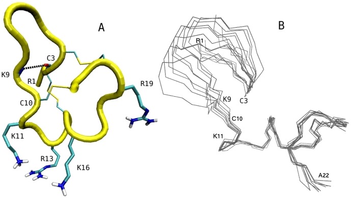
Figure 2. NMR structures of µ-GIIIA.
(A) Structure of µ-GIIIA with the pore blocking residues K11, R13 and K16 pointing downward. Three disulfide bridges and the C3–K10 hydrogen bond stabilizing the structure are indicated explicitly. (B) Superposition of the ten NMR structures demonstrating the flexibility of the N-terminal residues 1–9 around the C3 and C10 hinges.

