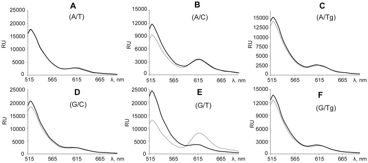
Figure 4. Fluorescence emission spectra.
Panels A–F correspond to DNA duplexes V–X in the presence of MutS (400 nM per monomer – dashed line) or in the absence of protein (solid line). DNA duplexes (concentration 20 nM) contain FRET pair - Alexa-488 (donor) and Alexa-594 (acceptor). The central variable nucleotide pair in DNA is shown in parentheses. The samples were irradiated by light at 470 nm. Spectra were recorded at 500-800 nm. RU - the signal detector in stated units. Each spectrum was recorded at least three times. The figure shows one of the experiments.

