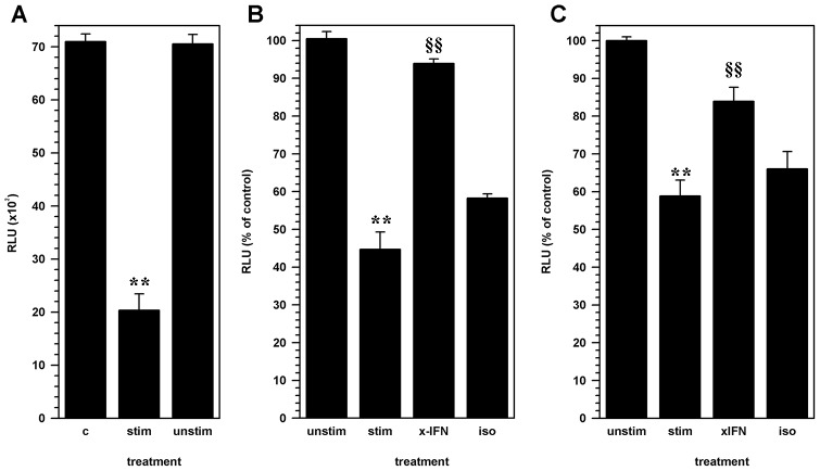
Figure 2. The effect of CD8 lymphocytes on NSC proliferation is mediated through IFN-γ.
Luc+ NSC cultures were treated with cell-free supernatants from activated CD8 lymphocytes 24 h after seeding cultures. (A) Luciferase activity measured from untreated NSCs (C) was compared to cultures treated with conditioned medium from antibody-activated CD8 lymphocyte (stim) or unstimulated CD8 lymphocytes (unstim). Pooled data from two separate experiments are presented. (B) Neutralizing antibodies to IFN-γ (x-IFN; 1 mg/ml) or an isotype control antibody (iso) was added to the activated-lymphocyte conditioned media prior to treatment of NSCs. Relative luminescence from NSC cultures treated with conditioned medium from unstimulated lymphocytes (unstim) or activated CD8 cells (stim) and treated activated-lymphocyte conditioned media are presented as a percent of untreated cultures. Pooled data from 3 independent experiments are presented. (C) NSC-lymphocyte co-cultures constituted with antibody-stimulated CD8 T-cells (stim) were treated with either neutralizing anti-IFN-γ antibody (xIFN), or an isotype antibody (iso). Relative luminescence intensity (RLU) from treated co-cultures and co-cultures with unstimulated CD8 T-cells (unstim) are presented as a percentage of untreated control NSCs. Pooled data from 3 independent experiments are presented (mean ± SEM; ** p<0.001 vs. untreated NSC; §§ p<0.001 vs. stimulated CD8 T-cell-NSC co-culture).

