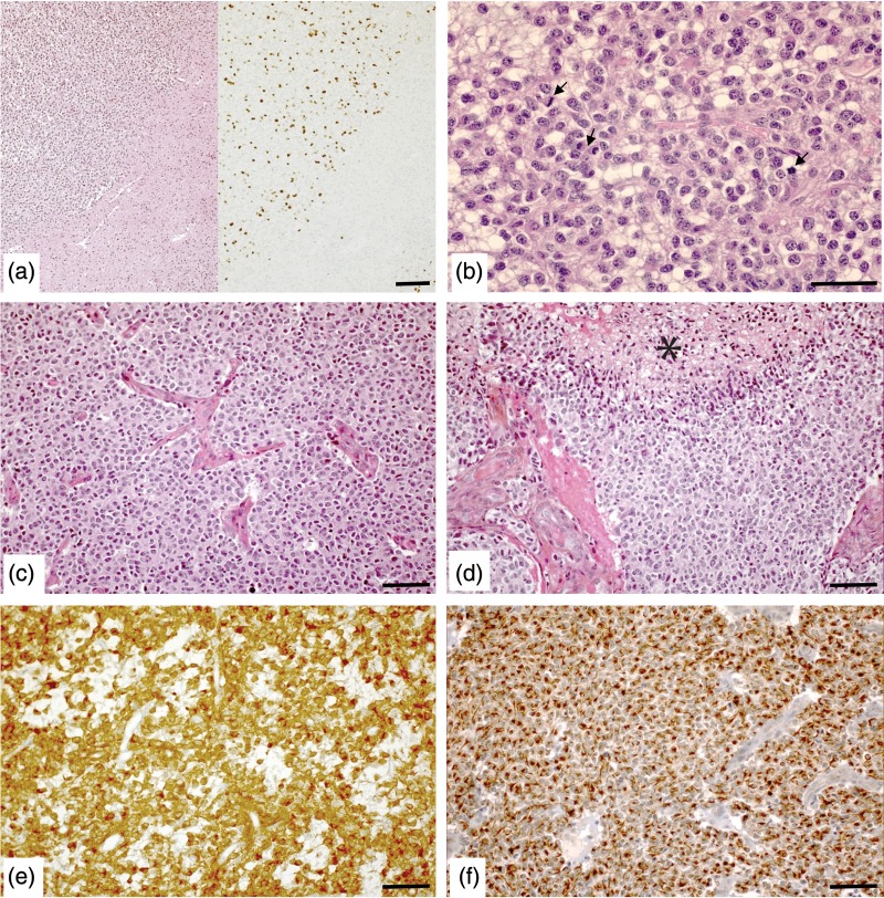
Fig. 1.
Pathological features of the 3 subgroups of anaplastic oligodendrogliomas. (a–c) Anaplastic oligodendroglioma with high mitotic count but no microvascular proliferation (MVP) and no palisading necrosis (PN). (a) Nodule of high cell density (hematoxylin-eosin [HE], left) with a KI 67 labeling index of 12% (immunostaining, right). (b) Higher magnification showing 3 mitotic figures (arrows) within one HPF (HE). (c) Anaplastic oligodendroglioma characterized by MVP (HE). (d) Anaplastic oligodendroglioma characterized by MVP and PN (*, HE). (e and f) Characteristic pattern of IDH1R132H (e) and internexin alpha (f) immunostaining in the same case recorded in panel a and b. Scale bar = 50 µm.

