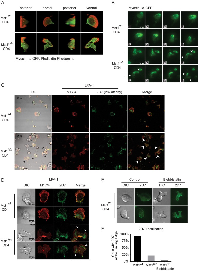
Figure 4. Mst1 regulates Myosin IIa localization and is required for partitioning of low and higher affinity LFA-1 molecules.
A) Wt and Mst1h/h CD4 T cells expressing Myosin IIa-GFP were seeded into slide chambers pre-coated with 1 μg/mL ICAM-1-Fc and stimulated with CCL19 prior to fixation and staining of F-actin with Rhodamine-phalloidin. Three-dimensional image reconstructions from z-stacks of confocal micrographs are displayed. B) Wt and Mst1h/h CD4 T cells expressing Myosin IIa-GFP were visualized by live TIRF microscopy. Arrows indicate bipolar morphology. Data are representative of 2 individual experiments with 150 cells per genotype. C, D) Wt and Mst1h/h CD4 T cells were stimulated as above and stained with 2D7 (anti-low affinity CD11a/LFA-1, green) and M17/4 (anti-CD11a/LFA-1, red). E) Wt CD4 T cells stimulated as above with or without Blebbistatin treatment were stained with 2D7 and visualized by immunofluorescence. F) Quantification of cells with 2D7 localization at the trailing edge of untreated or Blebbistatin-treated Wt and Mst1h/h CD4 T cells (data are representative of 2–3 individual experiments, n = 13–23).

