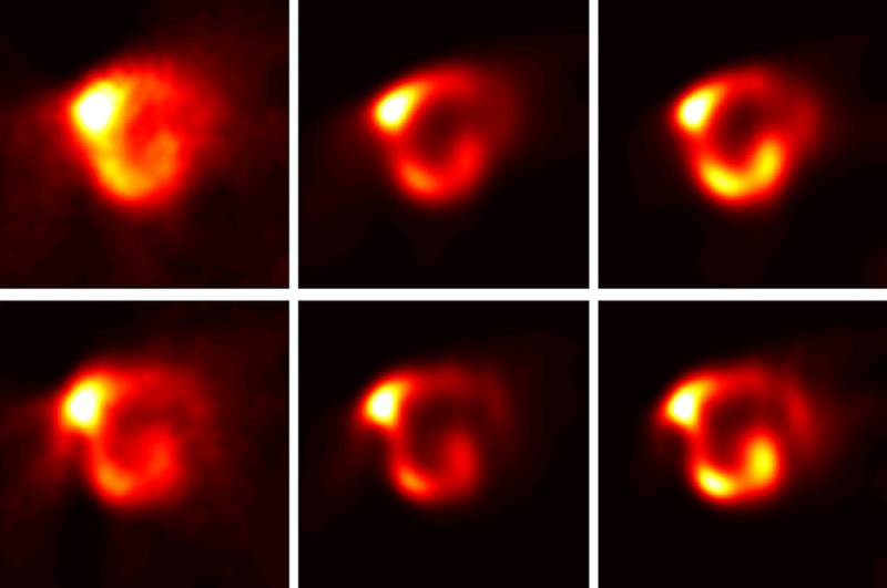
Figure 2.

The zoomed transaxial slices of SPECT images pertaining to the same patient are presented. The reconstructions with different time points upper row 9 h after therapy and lower row 20 h after therapy. First column: no correction, second column correction for bremsstrahlung and resolution loss, and third column correction for bremsstrahlung, resolution loss, attenuation and scatter correction as well. There is limited corrections (columns 1 and 2) compare to all corrections (column 3). The most advanced reconstruction technique resulted in high-quality representation of the biodistribution of microspheres in spite of the low abundance (15%) of the 188Re photopeak and high bremsstrahlung background.
