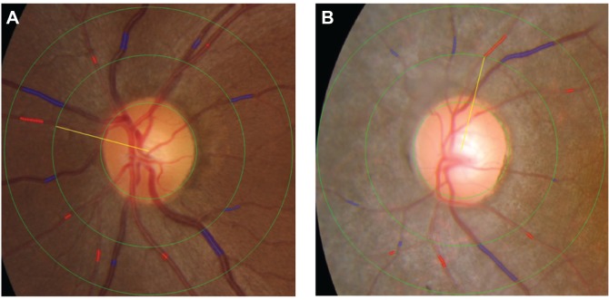
Figure 1.
Identification and measurement of the retinal arteriole and venule caliber using the IVAN semi-automated computer imaging program.
Notes: The blue and red shading indicates the selected venule and arteriole area, respectively, used to determine vessel caliber. (A) Color photograph of the left eye of a 32-year-old woman in the control group. More than six venules and arterioles could easily be measured in all normal patients. (B) Color photograph of the left eye of a 22-year-old woman with retinitis pigmentosa. Seven venules and only five arterioles could be measured.
Abbreviation: IVAN, interactive vessel analysis.

