Figure 3.

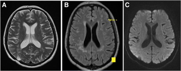
MRI-scan at 1-year follow-up visit. Markedly reduced white matter lesions. Right occipital hyperintensity on T2 reflects brain biopsy area. A) T2, B) T2-FLAIR, C) DWI.

MRI-scan at 1-year follow-up visit. Markedly reduced white matter lesions. Right occipital hyperintensity on T2 reflects brain biopsy area. A) T2, B) T2-FLAIR, C) DWI.