Figure 5.
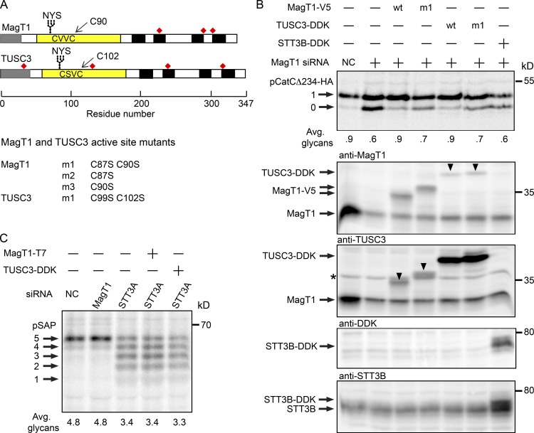
Requirement for active site cysteine residues in MagT1 and TUSC3. (A) Diagrams of MagT1 and TUSC3 showing the N-terminal signal sequence (gray), lumenal thioredoxin domain (yellow) with active-site CXXC motif, glycosylation site, noncatalytic cysteine residues (red squares), and membrane spanning segments (black). Nomenclature for single and double cysteine mutants of MagT1 and TUSC3 is given. (B) HeLa cells were treated with NC or MagT1 siRNA for 48 h before cotransfection with a pCatCΔ234-HA expression vector and a wild-type or mutant MagT1-V5, TUSC3-DDK, or STT3B-DDK expression vector. Cells were pulse labeled for 4 min and chased for 10 min. Glycoforms of CatCΔ234-HA were collected by immunoprecipitation with anti-HA and resolved by PAGE in SDS. Total protein extracts from cells were resolved by SDS-PAGE and analyzed by protein immunoblotting using anti-MagT1, anti-TUSC3, anti-DDK, and anti-STT3B sera. Downward pointing arrowheads in the anti-MagT1 and anti-TUSC3 blots designate cross-reaction with TUSC3-DDK and MagT1-V5, respectively. The band designated by the asterisk is not TUSC3, but is a nonspecific background protein (see Fig. S2 B). (C) HeLa cells treated for 48 h with NC, STT3A siRNA, or MagT1 siRNA were transfected with MagT1-T7 or TUSC3-DDK expression vectors as indicated 24 h before pulse labeling for 4 min and chase for 10 min. Prosaposin glycoforms were immunoprecipitated using anti–saposin D sera and resolved by SDS-PAGE. Quantified values below gel lanes (B and C) are for the displayed image that is representative of two experiments.

