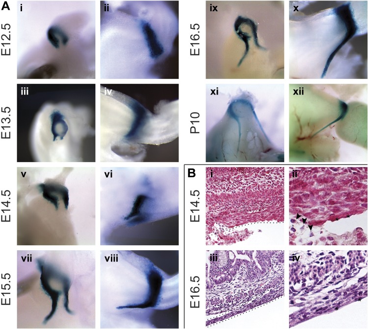
Fig. 1.
Morphogenesis and composition of the murine gastric ligaments. A: whole mount X-gal staining of Gata3lacZ/+ embryos. i and ii: The gastric ligaments have not yet developed at embryonic day (E) 12.5, although Gata3 is expressed in a C-shaped expression domain at the pylorus. The open portion of the “C” is ventral, which produces a Gata3 expression gap. iii and iv: At E13.5, bilateral Gata3-positive cellular cords (i.e., the nascent gastric ligaments) begin to extend anteriorly from either edge of the ventral Gata3 pyloric expression gap. v–x: From E14.5 to E16.5, the nascent gastric ligaments extend superficially and anteriorly over the ventral (lesser curvature) side of the stomach to reach the level of the esophagus. xi and xii: The gastric ligaments are still visible at postnatal day (P) 10. B: hematoxylin and eosin staining of wild-type (WT) embryos. i and ii: At E14.5, round-to-oval, mesenchymal-type cells compose the core of the ligaments; the ligament surface is lined by flattened, squamous-type serosal cells (asterisks) and anchored to the ventral stomach by loose connective tissue (arrowheads). iii and iv: By E16.5, the gastric ligaments have elongated and thinned but maintained their overall architecture. The cells of the ligament cores have adopted a spindle-shaped, smooth muscle-type morphology, with cellular and nuclear elongation parallel to the anteroposterior (long) axis of the gastrointestinal tract lumen. Orientation is as follows: stomach is left, and dorsal is up.

